Alfalfa:Photos
Alfalfa Pest and Nutritional Disorder Photos
Contents
- 1 Agrobacterium_tumefaciens
- 2 Alfalfa_mosaic_virus
- 3 Alfalfa_mosaic_virus
- 4 Alfalfa_mosaic_virus
- 5 Alfalfa_stem_nematode_2
- 6 Alfalfa_stem_nematode_damage
- 7 Alfalfa_stem_nematode_damage
- 8 Anthracnose_-_Sorghum
- 9 Aphanomyces_root_rot
- 10 Ascochyta_Rough_Spot_-_Sorghum
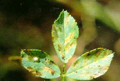
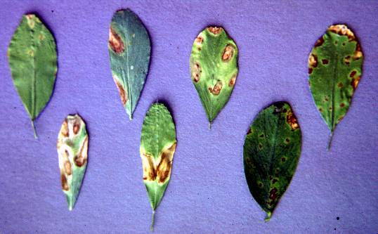
- 11 Bacterial_leaf_spot
- 12 Bacterial_leaf_spot
- 13 Bacterial_leaf_spot_1
- 14 Bacterial_leaf_spot_2
- 15 Bacterial_leaf_spot_3
- 16 Bacterial_wilt
- 17 Bacterial_wilt_symptoms
- 18 Bacterial_wilt_symptoms
- 19 Blister_beetle_on_alfalfa
- 20 Calonectria_crotalariae
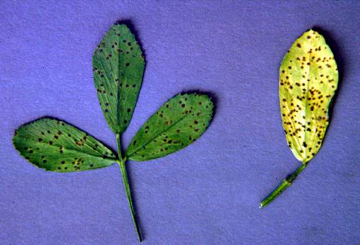
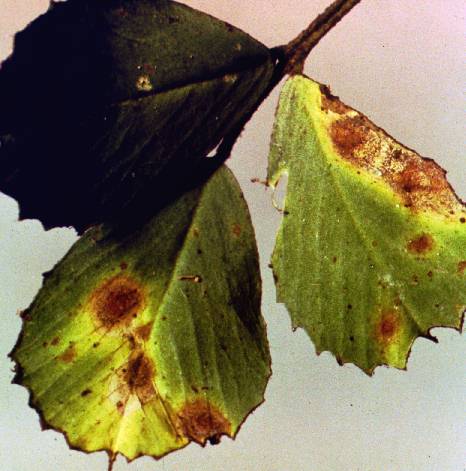
- 21 Cercospora_leaf_spot
- 22 Cercospora_on_leaves
- 23 Cercospora_on_stems
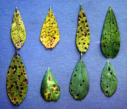
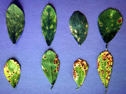
- 24 Common_leaf_spot
- 25 Common_leaf_spot
- 26 Common_leaf_spot_1
- 27 Common_leaf_spot_3
- 28 Dodder
- 29 Downy_mildew
- 30 Downy_mildew_1
- 31 Downy_mildew_2
- 32 Downy_mildew_3
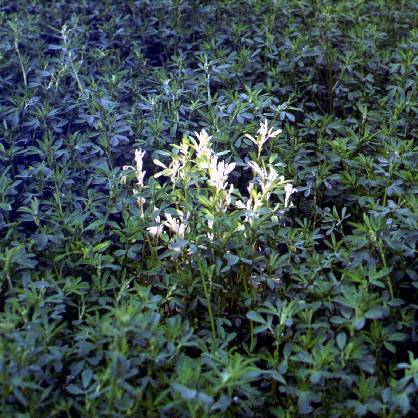
- 33 Downy_mildew_in_the_field
- 34 Foliar_tolerance
- 35 Frost_injury
- 36 Frost_injury
- 37 Fusarium_wilt
- 38 Fusarium_wilt
- 39 Fusarium_wilt_(left)_and_bacterial_wilt_(right)
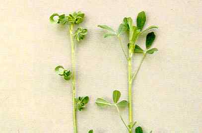
- 40 Genetic_abnormailty_12
- 41 Genetic_abnormailty_15
- 42 Genetic_abnormality_00
- 43 Genetic_abnormality_01
- 44 Genetic_abnormality_02
- 45 Genetic_abnormality_03
- 46 Genetic_abnormality_04
- 47 Genetic_abnormality_05
- 48 Genetic_abnormality_06
- 49 Genetic_abnormality_07
- 50 Genetic_abnormality_08
- 51 Genetic_abnormality_09
- 52 Genetic_abnormality_10
- 53 Genetic_abnormality_11
- 54 Genetic_abnormality_13
- 55 Genetic_abnormality_14
- 56 Helminthosporium_Leaf_Spot_-_Bermuda
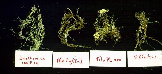
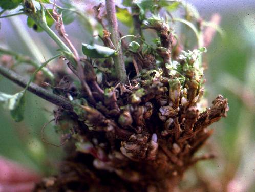
- 57 Ineffective_nodulation
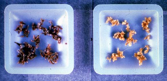
- 58 Ineffective_nodules
- 59 Leaf_Spot_-_Alfalfa_Pseudopeziza_Jonesii
- 60 Leaf_Spot_-_Alfalfa_Pseudopeziza_Medicaginis
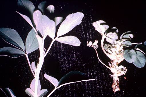
- 61 Lepto_leaf_spot
- 62 Neocosmospora_vasinfecta
- 63 Nitrogen_deficiency
- 64 Nitrogen_deficiency
- 65 Ozone_damage
- 66 Peanut_Mottle_Virus_-_Clover
- 67 Phytophthora_medicaginis
- 68 Phytophthora_medicaginis
- 69 Phytophthora_root_rot
- 70 Phytophthora_root_rot
- 71 Phytophthora_root_rot
- 72 Pseudoplea_Leaf_Spot_-_Clover
- 73 Rhizobium_nodules_1
- 74 Rhizoctonia
- 75 Rhizoctonia_symptoms
- 76 Root_knot_nematode
- 77 Root_knot_nematode
- 78 Root_knot_nematode_symptoms
- 79 Root-lesion_nematode
- 80 Root-lesion_nematode
- 81 Root-lesion_nematodes
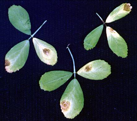
- 82 Rust_2
- 83 Rust_symptoms
- 84 Rust_symptoms
- 85 Sclerotium_1
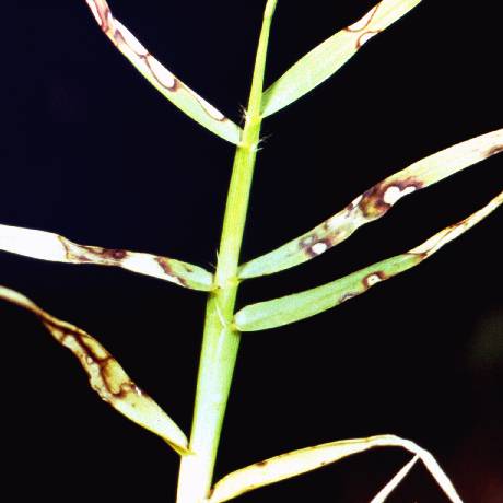
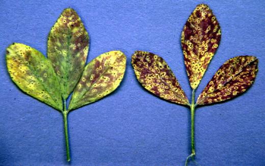
- 86 Spring_blackstem_and_leaf_spot
- 87 Spring_blackstem_and_leaf_spot
- 88 Stagnospora_symptoms
- 89 Stagnospora_symptoms
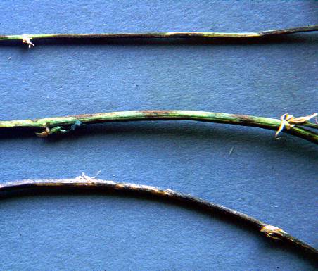
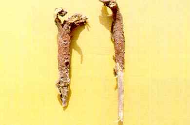
- 90 Stem_canker
- 91 Stem_nematode
- 92 Stem_nematode
- 93 Stem_nematode
- 94 Stem_nematode
- 95 Stem_nematode_on_inoculated_seedlings
- 96 Stemphylium_symptoms
- 97 Sulfur_dioxide_injury
- 98 Tarspot
- 99 Verticillium_albo-atrum_1
- 100 Verticillium_albo-atrum_3
- 101 Verticillium_albo-atrum_4
- 102 Verticillium_wilt
- 103 Verticillium_wilt
- 104 Verticillium_wilt
- 105 Verticillium_wilt
- 106 Violet_root_rot_2
- 107 Yellow_leaf_blotch_1
Agrobacterium_tumefaciens
Agrobacterium tumefaciens Large galls from inoculation of young plants with Agrobacterium tumafaciens (Deborah Samac)
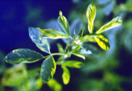
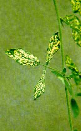
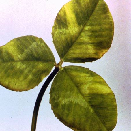
Alfalfa_mosaic_virus
Alfalfa mosaic virus (Fred Frosheiser)
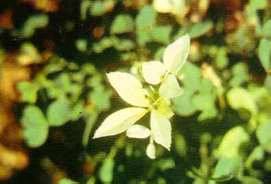
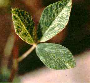
Alfalfa_mosaic_virus
Alfalfa mosaic virus (Fred Frosheiser)
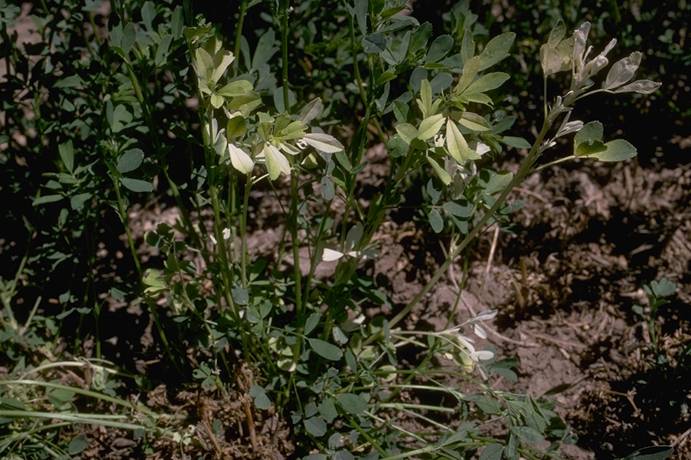
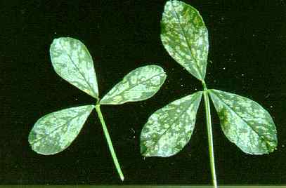
Alfalfa_mosaic_virus
Alfalfa mosaic virus (Fred Frosheiser)
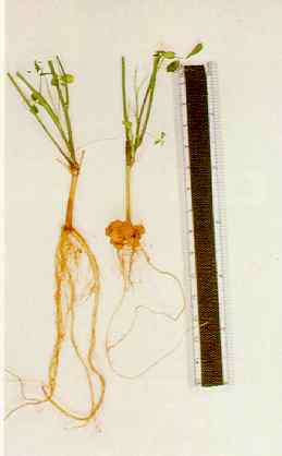
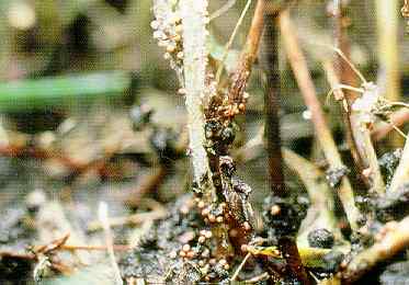
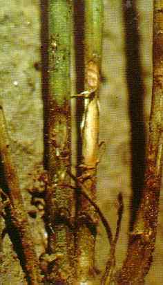
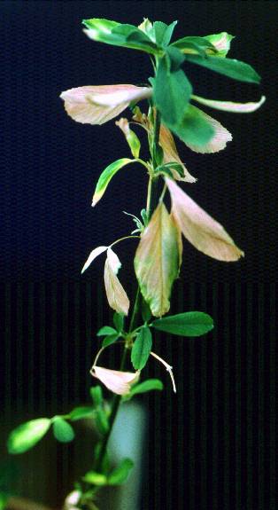
Alfalfa_stem_nematode_2
Alfalfa stem nematode white flagging, Ditylenchus dipsaci (Gerald D. Griffin, USDA)
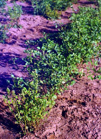
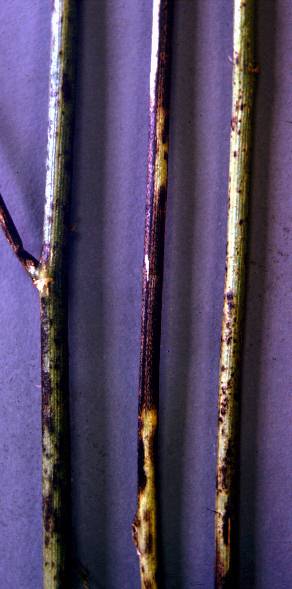
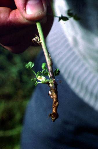
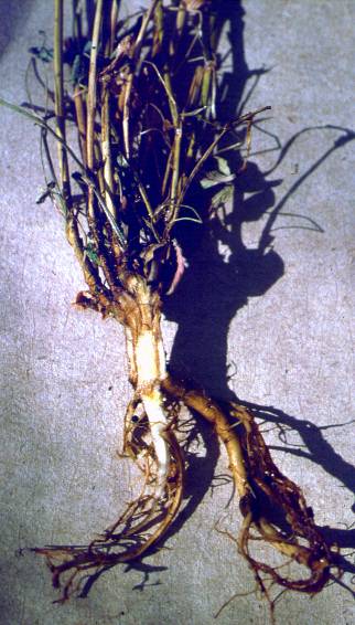
Alfalfa_stem_nematode_damage
Alfalfa stem nematode damage (H. F. Schwartz; Colorado State University)

Alfalfa_stem_nematode_damage
Alfalfa stem nematode damage (H. F. Schwartz; Colorado State University)
Anthracnose_-_Sorghum
Anthracnose - Sorghum
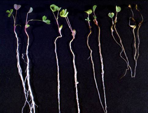
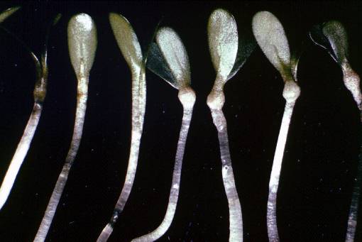
Aphanomyces_root_rot
Aphanomyces root rot on inoculated seedlings; rating 1 (healthy) to 5 (dead) (Deborah Samac)
Ascochyta_Rough_Spot_-_Sorghum
Ascochyta Rough Spot - Sorghum
Bacterial_leaf_spot
Bacterial leaf spot (Fred Frosheiser)
Bacterial_leaf_spot
Bacterial leaf spot (Fred Frosheiser)
Bacterial_leaf_spot_1
Bacterial leaf spot water-soaked lesions (Donald L. Stuteville, Kansas State University)
Bacterial_leaf_spot_2
Bacterial leaf spot translucent lesions (Donald L. Stuteville, Kansas State University)
Bacterial_leaf_spot_3
Bacterial leaf spot lesions, alfalfa stems 3 weeks after inoculation (Donald L. Stuteville, Kansas State University
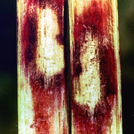
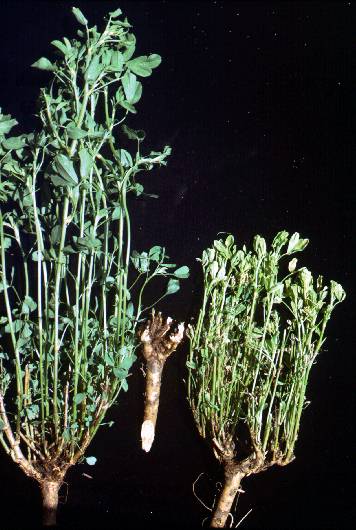
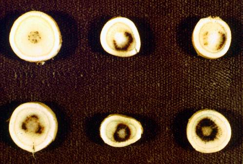
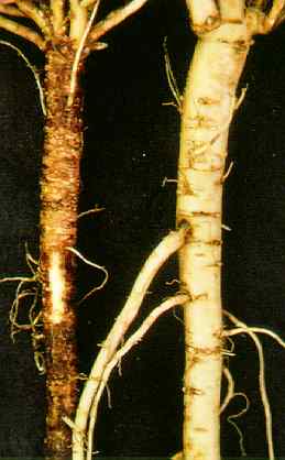
Bacterial_wilt
Bacterial wilt root cross sections. Most diseased (top left) to healthy (bottom right) (Barnes)
Bacterial_wilt_symptoms
Bacterial wilt symptoms in the field (front plant) compared to a healthy plant behind (Deborah Samac)
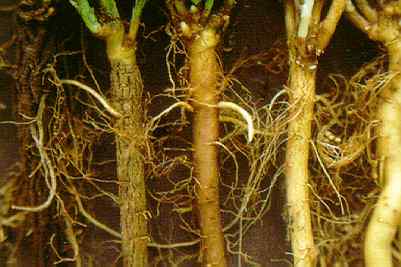
Bacterial_wilt_symptoms
Bacterial wilt symptoms. Healthy plant (left), diseased root section (middle), diseased plant (right) (Barnes)
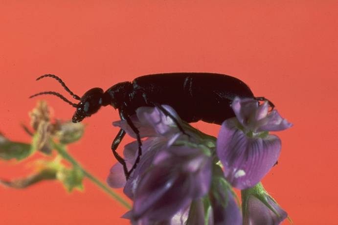
Blister_beetle_on_alfalfa
Blister beetle on alfalfa (Colorado State University)
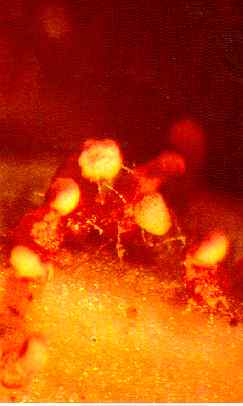
Calonectria_crotalariae
Calonectria crotalariae infected alfalfa plant (base and stem) (Donald L. Stuteville, Kansas State University)
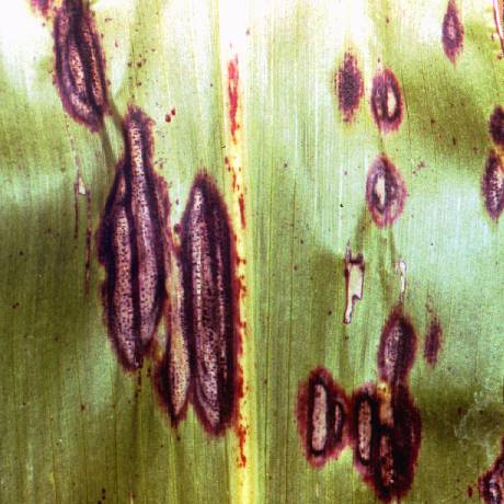
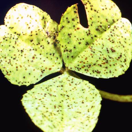
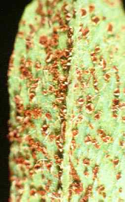
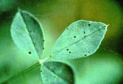
Cercospora_leaf_spot
Leaf spot, Cercospora medicaginis (William G. Willis, Kansas State University)

Cercospora_on_leaves
Cercospora on leaves (Fred Frosheiser)
Cercospora_on_stems
Cercospora on stems (Fred Frosheiser)
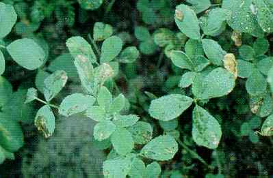
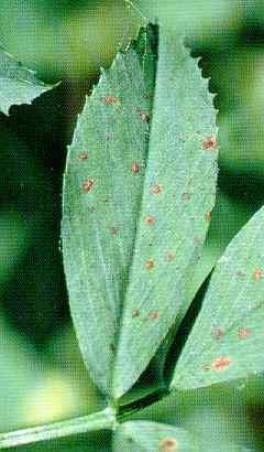
Common_leaf_spot
Common leaf spot (Fred Frosheiser)
Common_leaf_spot
Commonleaf spot (Fred Frosheiser)
Common_leaf_spot_1
'Common leaf spot (William G. Willis, Kansas State University)
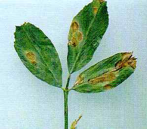
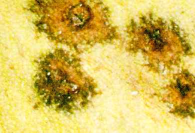
Common_leaf_spot_3
Common leaf spot lesions (magnified view), dentate margins, Pseudopeziza medicaginis apothecia rupturing, host epidermis (William G. Willis, Kansas State University)
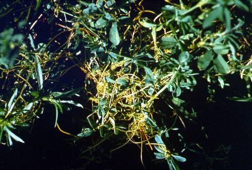
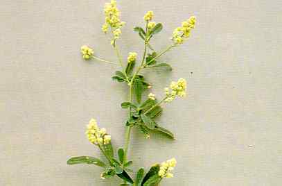
Dodder
Dodder (USDA-ARS)
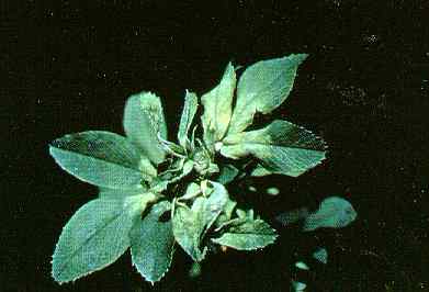
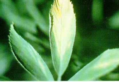
Downy_mildew
Downy mildew (Fred Frosheiser)
Downy_mildew_1
Shoot systemically infected with downy mildew fungus, Peronospora trifoliorum (Kenneth T. Leath, USDA)
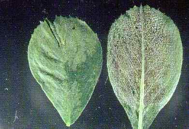
Downy_mildew_2
Downy mildew, upper (left), lower (right) surfaces, leaflets (Donald L. Stuteville, Kansas State University)
Downy_mildew_3
Downy mildew fungus oospores (Peronospora trifoliorum), cotyledon stained, sudan iv (Donald L. Stuteville, Kansas State University)
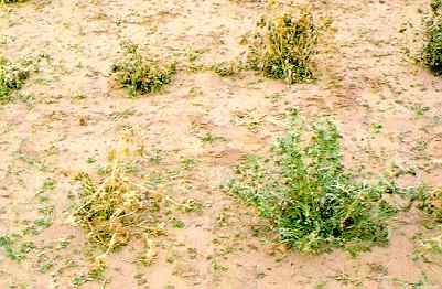
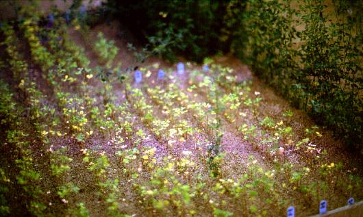
Downy_mildew_in_the_field
Downy mildew in the field (Fred Frosheiser)

Foliar_tolerance
Differences in foliar tolerance, low temperature among spaced plants (Donald L. Stuteville, Kansas State University)
Frost_injury
Frost injury (Barnes)
Frost_injury
Frost injury (Barnes)
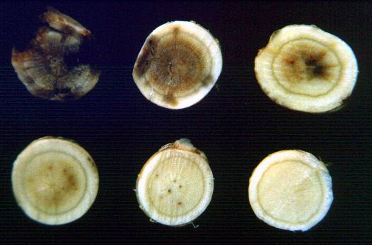
Fusarium_wilt
Fusarium wilt in alfalfa roots, cross sections. Healthy (top left) to most diseased (bottom right) (Fred Frosheiser)
Fusarium_wilt
Fusarium wilt in alfalfa roots, longitudinal section (Barnes)
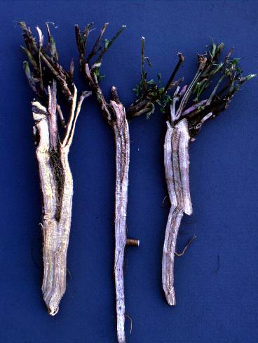
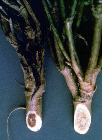
Fusarium_wilt_(left)_and_bacterial_wilt_(right)
Fusarium wilt (left) and bacterial wilt (right) (Barnes)
Genetic_abnormailty_12
Genetic abnormality: taproot tumors (Donald K. Barnes, USDA)
Genetic_abnormailty_15
Genetic abnormality: heavy bark condition, rough root showing lack, functional small roots (Donald K. Barnes, USDA)
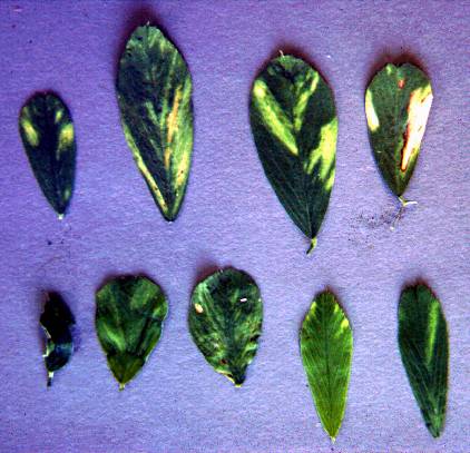
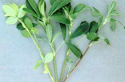
Genetic_abnormality_00
Genetic abnormalities : pale green leaves, stem (left), normal green leaves, stem, light red stem, dark red stem (right)(Donald K. Barnes, USDA)
Genetic_abnormality_01
Genetic abnormality : white leaf spotting induced, low temperatures, long days (Donald K. Barnes, USDA)
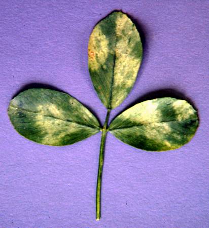
Genetic_abnormality_02
Genetic abnormality : mottled leaf (Donald K. Barnes, USDA)
Genetic_abnormality_03
Genetic abnormality : white mosaic leaf spotting (Donald K. Barnes, USDA)
Genetic_abnormality_04
Genetic abnormality : golden tip, characterized, chlorosis, newly developed leaves (Donald K. Barnes, USDA)
Genetic_abnormality_05
Genetic abnormality : curled leaf (Donald K. Barnes, USDA)
Genetic_abnormality_06
Genetic abnormality : vestigial flower, branched raceme (Donald K. Barnes, USDA)
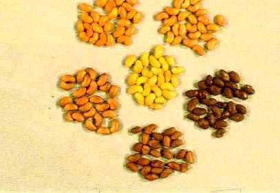
Genetic_abnormality_07
Genetic variability , seed color: normal (center), tan, black, dark red, mottled, light red mottled seed colors, nonuniform distribution, red endosperm pigment (Donald K. Barnes, USDA)
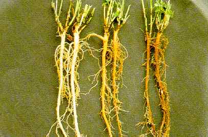
Genetic_abnormality_08
Genetic abnormalities white (normal), tan, brown roots (Donald K. Barnes, USDA)
Genetic_abnormality_09
Genetic abnormality : red root (Donald K. Barnes, USDA)

Genetic_abnormality_10
Genetic abnormality : cross section, roots showing normal white internal root (left), orange root (Donald K. Barnes, USDA)

Genetic_abnormality_11
Genetic abnormality : stem cuttings showing normal rooting (left), abnormal callus tumor (Donald K. Barnes, USDA)
Genetic_abnormality_13
Genetic abnormality : excessive root lenticel development, healthy tissue underneath (left), normal root, plant grown, wet soil (Donald K. Barnes, USDA)
Genetic_abnormality_14
Genetic abnormality : variability, expression, rough root; normal root, right (Donald K. Barnes, USDA)
Helminthosporium_Leaf_Spot_-_Bermuda
Helminthosporium Leaf Spot - Bermuda
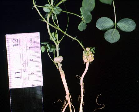
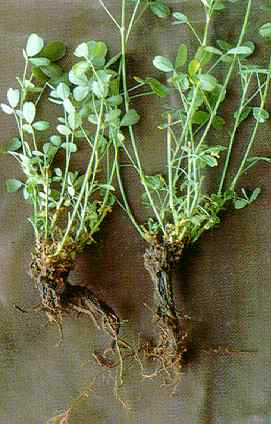
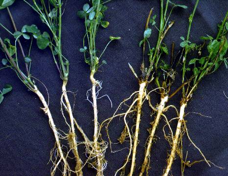
Ineffective_nodulation
Ineffective nodulation (2 plants on left), effective nodulation (2 plants on right) (Barnes)
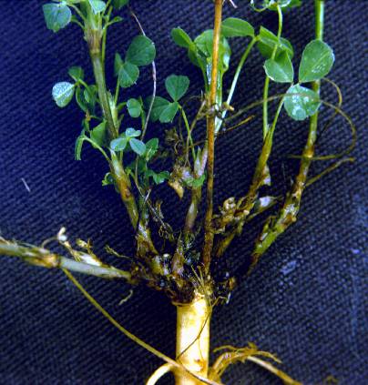
Ineffective_nodules
White ineffective nodules (right) and effective nodules (left) (Barnes)
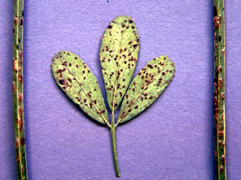
Leaf_Spot_-_Alfalfa_Pseudopeziza_Jonesii
Leaf Spot - Alfalfa Pseudopeziza Jonesii
Leaf_Spot_-_Alfalfa_Pseudopeziza_Medicaginis
Leaf Spot - Alfalfa Pseudopeziza Medicaginis

Lepto_leaf_spot
Lepto leaf spot (Fred Frosheiser)
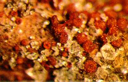
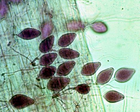
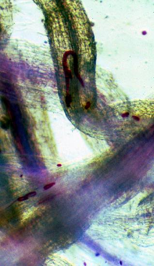
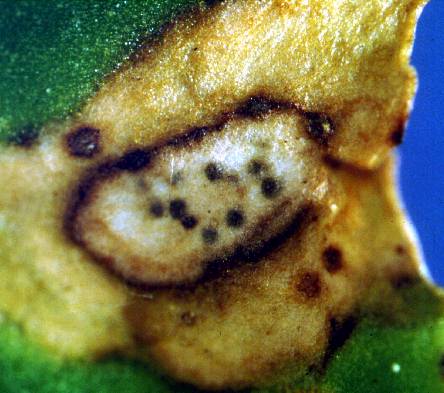
Neocosmospora_vasinfecta
Neocosmospora vasinfecta perithecia (red) exuding ascospores (orange) on alfalfa stem, water agar (Donald L. Stuteville, Kansas State University)
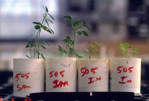
Nitrogen_deficiency
Ineffective plants growing in sand with nil nitrogen (left) compared to effective plants (right) (Barnes)
Nitrogen_deficiency
Ineffective plants growing in sand with nil nitrogen (right) compared to effective plants (left) (Barnes)
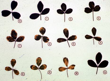
Ozone_damage
Ozone damage rated 0 (no damage) to 10 (severe damage) (Fred Frosheiser)
Peanut_Mottle_Virus_-_Clover
Peanut Mottle Virus - Clover
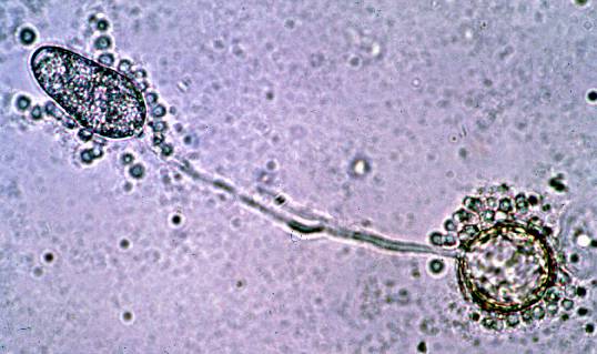
Phytophthora_medicaginis
Germinating oospore of Phytophthora medicaginis with zoosporangium (Fred Frosheiser)
Phytophthora_medicaginis
Zoosporangia of Phytophthora medicaginis stained red (Fred Frosheiser)
Phytophthora_root_rot
Phytophthora root rot symptoms in July (Fred Frosheiser)

Phytophthora_root_rot
Phytophthora root rot, healthy (left) to most diseased (right) (Barnes)
Phytophthora_root_rot
Phytophthora root rot, young plants (Fred Frosheiser)

Pseudoplea_Leaf_Spot_-_Clover
Pseudoplea Leaf Spot - Clover
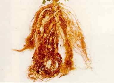
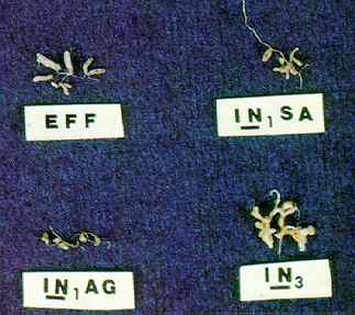
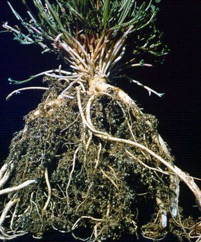
Rhizobium_nodules_1
Effective (eff) rhizobium nodules on alfalfa root, three types, ineffective (in) nodules induced, alfalfa plant genotype (Carroll P. Vance, USDA)
Rhizoctonia
Rhizoctonia (3 plants on right), healthy (3 plants on left) (Barnes)
Rhizoctonia_symptoms
Rhizoctonia symptoms (Barnes)
Root_knot_nematode
Root knot nematode symptoms (left and right), healthy plant (middle) (Fred Frosheiser)

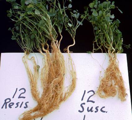
Root_knot_nematode
Root knot nematode 12 healthy plants (left), 12 susceptible plants (right) (Fred Frosheiser)
Root_knot_nematode_symptoms
Root knot nematode symptoms (Fred Frosheiser)
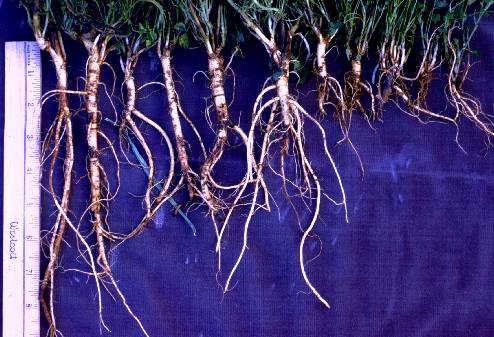
Root-lesion_nematode
Root-lesion nematode field symptoms (center), healthy plants (left and right) (Deborah Samac)
Root-lesion_nematode
Root-lesion nematode symptoms (right) and healthy plant (left) (Theis)
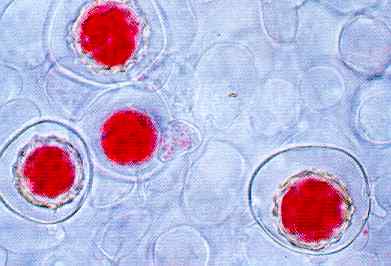
Root-lesion_nematodes
Root-lesion nematodes and eggs stained red in alfalfa roots (Deborah Samac)
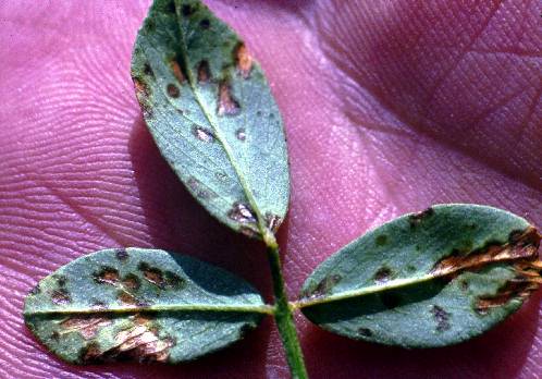
Rust_2
Rust on leaflet, uredinial pustules (Donald L. Stuteville, Kansas State University)
Rust_symptoms
Rust symptoms (Fred Frosheiser)
Rust_symptoms
Rust symptoms and potato leaf hopper symptoms (Fred Frosheiser)
Sclerotium_1
Sclerotim Mature (brown) sclerotia (Sclerotium rolfsii) on plant base, Mycelium producing sclerotia progresses up the stem (Donald L. Stuteville, Kansas State University)
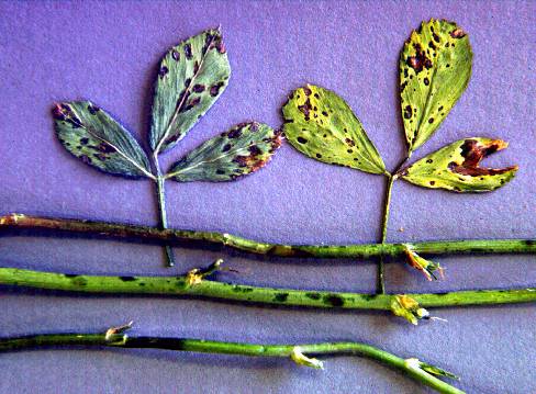
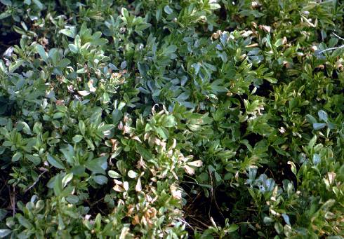
Spring_blackstem_and_leaf_spot
Spring blackstem and leaf spot (Fred Frosheiser)

Spring_blackstem_and_leaf_spot
Spring blackstem and leaf spot (Fred Frosheiser)
Stagnospora_symptoms
Stagnospora symptoms on leaf (Fred Frosheiser)
Stagnospora_symptoms
Stagnospora symptoms on the crown (Fred Frosheiser)
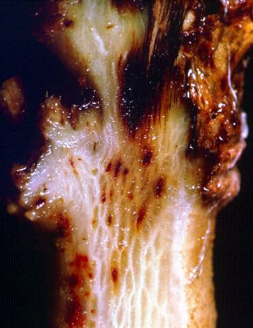
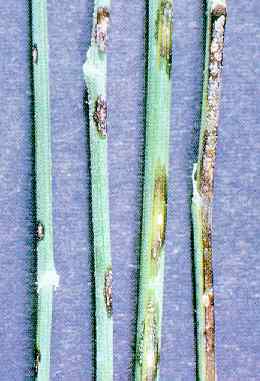
Stem_canker
Stem canker, Rhizoctonia solani (William G. Willis, Kansas State University)
Stem_nematode
Stem nematode (Fred Frosheiser)
Stem_nematode
Stem nematode (Fred Frosheiser)
Stem_nematode
Stem nematode (Fred Frosheiser)
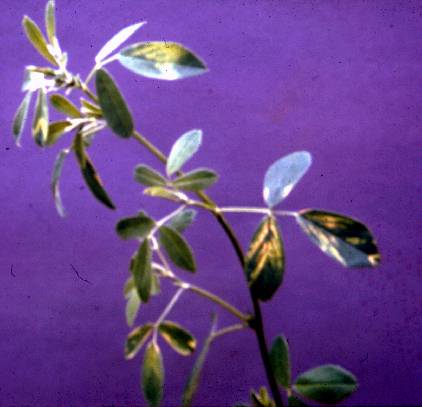
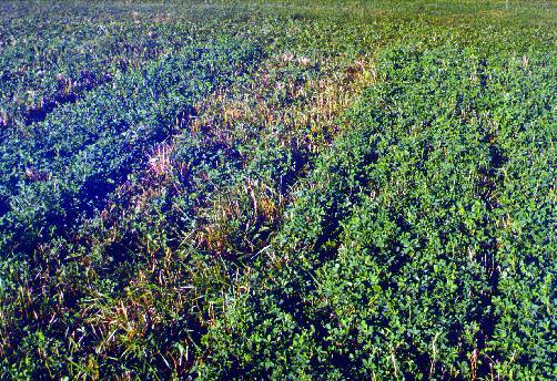
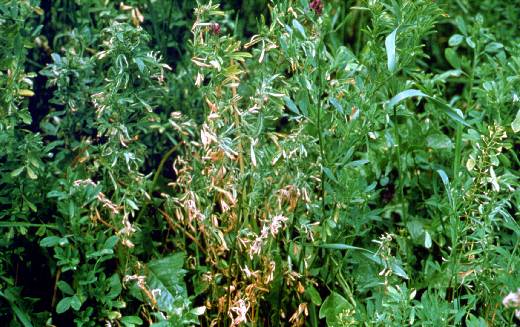
Stem_nematode
Stem nematode symptoms in the field (white flagging) (Fred Frosheisegr)
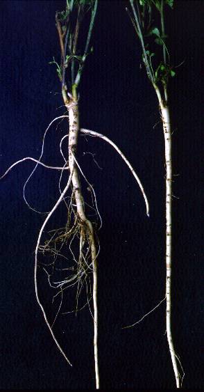
Stem_nematode_on_inoculated_seedlings
Stem nematode on inoculated seedlings Resistant plants (left); susceptible plants (right) (Fred Forosheiser)
Stemphylium_symptoms
Stemphylium symptoms (Fred Frosheiser)
Sulfur_dioxide_injury
Sulfur dioxide injury (Kenneth T. Leath, USDA)
Tarspot
Tarspot, Phoma medicaginis (Kenneth T. Leath, USDA)
Verticillium_albo-atrum_1
Verticillium albo-atrum infected leaflets, v-shaped chlorosis, tip, curling (Kenneth T. Leath, USDA)
Verticillium_albo-atrum_3
Verticillium albo-atrum infected leaflets, pinkish necrosis (Kenneth T. Leath, USDA)
Verticillium_albo-atrum_4
Verticillium albo-atrum necrotic leaves infected (Kenneth T. Leath, USDA)
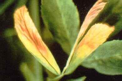
Verticillium_wilt
Verticillium wilt crown symptoms (Fred Frosheiser)
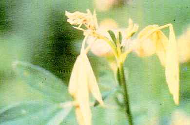
Verticillium_wilt
Verticillium wilt foliar symptoms (Fred Frosheiser)
Verticillium_wilt
Verticillium wilt foliar symptoms (Fred Frosheiser)

Verticillium_wilt
Verticillium wilt in the field (Fred Frosheiser)
Violet_root_rot_2
Violet root rot fungus (Rhizoctonia crocorum) on crown/root (William G. Willis, Kansas State University)
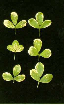
Yellow_leaf_blotch_1
Yellow leaf blotch early symptoms (William G. Willis, Kansas State University)