Grapes:Photos
Grape Pest and Nutritional Disorder Photos
Contents
- 1 Ajinashika_disease
- 2 AMV_infection
- 3 AMV_infection
- 4 AMV_infection
- 5 AMV_infection
- 6 AMV_Infection
- 7 AMV_infection
- 8 Anthracnose_Bird's_-_Eye_Rot
- 9 Anthracnose_Soft_Grape
- 10 ArMV_induced_mosaic_mottle
- 11 ArMV_infection
- 12 ArMV_infection
- 13 ArMV_infection
- 14 Asteroid_mosaic
- 15 Asteroid_mosaic_virus_(AMV)_infection
- 16 Bitter_Rot
- 17 Black_Rot_on_Fruit
- 18 Black_Rot_on_Leaf
- 19 Black_Rot_on_Stem
- 20 Bois_noir_infection
- 21 Bois_noir_infection
- 22 Bois_noir_infection
- 23 Bois_noir_infection
- 24 CarMV_infection
- 25 CarMV_infection
- 26 Chrome_mosaic
- 27 Chrome_mosaic_symptoms
- 28 Concord_grape
- 29 Corky_bark
- 30 Corky_bark
- 31 Corky_bark
- 32 Corky_bark
- 33 Corky_bark
- 34 Cristulariella_1
- 35 Downy_Mildew
- 36 Enations
- 37 Enations
- 38 Enations
- 39 Fanleaf_infection
- 40 Fanleaf_infection
- 41 Fanleaf_infection
- 42 Fanleaf_infection
- 43 Fanleaf_infection
- 44 Fanleaf_infection
- 45 Fanleaf_infection
- 46 Fanleaf_symptoms
- 47 Flavescence_doree_infection
- 48 Flavescence_doree_infection
- 49 Flavescence_doree_infection
- 50 Flavescence_doree_infection
- 51 Flavescence_doree_infection
- 52 Flavescence_doree_infection
- 53 Flavescence_doree_infection
- 54 Flavescence_doree_infection
- 55 Fleck_disease
- 56 GCMV_infection
- 57 GCMV_infection
- 58 GCMV_infection
- 59 GCMV_infection
- 60 GCMV_infection
- 61 GCMV_infection
- 62 GFLV_infection
- 63 GFLV_infection
- 64 GFLV_infection
- 65 GFLV_infection
- 66 GFLV_infection
- 67 GFLV_infection
- 68 GLPV_infection
- 69 GLPV_infection
- 70 GLPV_infection
- 71 Grapevine_leafroll-associated
- 72 Grapevine_stunt
- 73 GVA_infection
- 74 Isariopsis_Leaf_Spot
- 75 Leaf_blotch_1
- 76 Leaf_discoloration
- 77 Leaf_discoloration
- 78 Leafroll_infection
- 79 Leafroll_infection
- 80 Leafroll_infection
- 81 Leafroll_symptoms
- 82 Leafroll_symptoms
- 83 Leafroll_symptoms
- 84 Leafroll_symptoms
- 85 Leafroll_symptoms
- 86 Line_pattern_infection
- 87 Line_pattern_infection
- 88 Line_pattern_infection
- 89 Pierce's_disease_(Xylella_fastidiosa)
- 90 Pierce's_disease_(Xylella_fastidiosa)
- 91 Pierce's_disease_(Xylella_fastidiosa)
- 92 Pierce's_disease_(Xylella_fastidiosa)
- 93 Pierce's_disease_(Xylella_fastidiosa)
- 94 Pierce's_disease_(Xylella_fastidiosa)
- 95 Pierce's_disease_(Xylella_fastidiosa)
- 96 Pierce's_disease_(Xylella_fastidiosa)
- 97 Pierce's_disease_(Xylella_fastidiosa)
- 98 Pierce's_disease_(Xylella_fastidiosa)
- 99 Pierce's_disease_(Xylella_fastidiosa)
- 100 Pierce's_disease_(Xylella_fastidiosa)
- 101 Pierce's_disease_(Xylella_fastidiosa)
- 102 PRMV_infection
- 103 PRMV_infection
- 104 PRMV_infection
- 105 RRV_infection
- 106 Rugose_wood
- 107 Rugose_wood
- 108 Rugose_wood
- 109 Rugose_wood
- 110 Rugose_wood
- 111 Rugose_wood
- 112 Rugose_wood
- 113 Rugose_wood
- 114 Rugose_wood
- 115 Scaphoideus_titanus
- 116 SLRV_infection
- 117 SLRV_infection
- 118 SLRV_infection
- 119 Stem_grooving
- 120 Stem_grooving
- 121 Stem_pitting_disease
- 122 Stem_pitting_symptoms
- 123 Stunt_symptoms
- 124 Stunt_symptoms
- 125 Sulfur_dioxide_1
- 126 TBRV_infection
- 127 TBVR_infection
- 128 Tomato_ringspot_2
- 129 ToRsv_infection
- 130 ToRSV_infection
- 131 ToRSV_infection
- 132 ToRSV_infection
- 133 ToRSV_infection
- 134 TRSV_and_GFLV_infections
- 135 TRSV_infection
- 136 TRSV_infection
- 137 TRSV_infection
- 138 TRSV_infection
- 139 TRSV_infection
- 140 TRSV_infection
- 141 TRSV_infection
- 142 Vein_mosaic_disease
- 143 Vein_mosaic_disease
- 144 Vein_mosaic_disease
- 145 Vein_necrosis
- 146 Yellow_mosaic
- 147 Yellow_mosaic
- 148 Yellow_mosaic
- 149 Yellow_mosaic
- 150 Yellow_mosaic
- 151 Yellow_mosaic
- 152 Yellow_speckle_and_leafroll
- 153 Yellow_speckle_infection
- 154 Yellow_speckle_infection
- 155 Yellow_speckle_infection
- 156 Yellow_speckle_viroid_and_grapevine_fanleaf_virus
- 157 Yellow_speckle_viroid_and_grapevine_fanleaf_virus
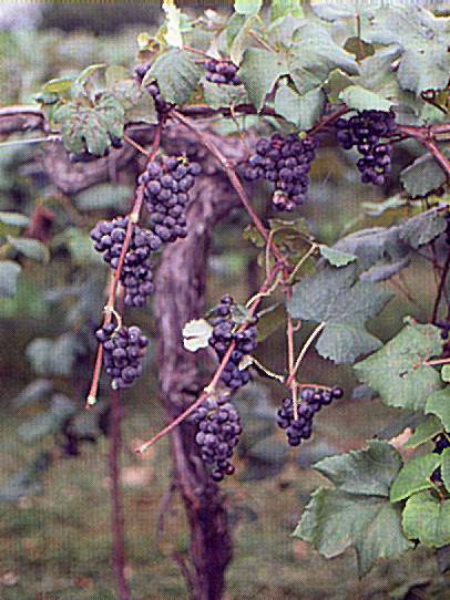
Ajinashika_disease
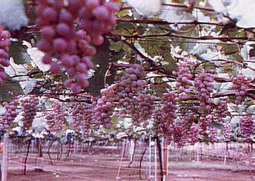
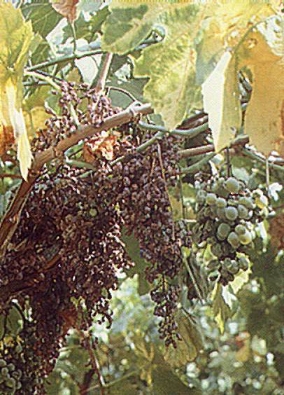
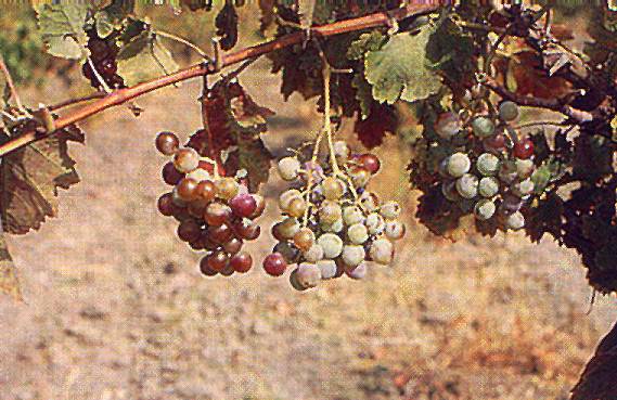
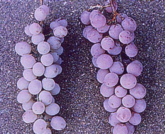
Ajinashika disease
A cv. Koshu vineyard with vines affected by ajinashika disease, showing pale-coloured bunches
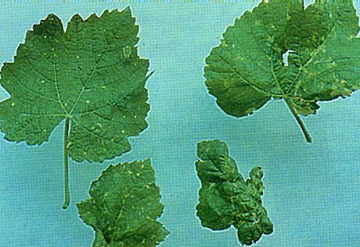
AMV_infection
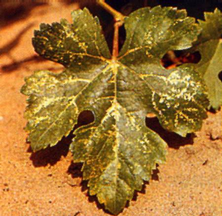
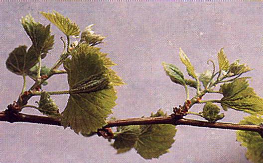
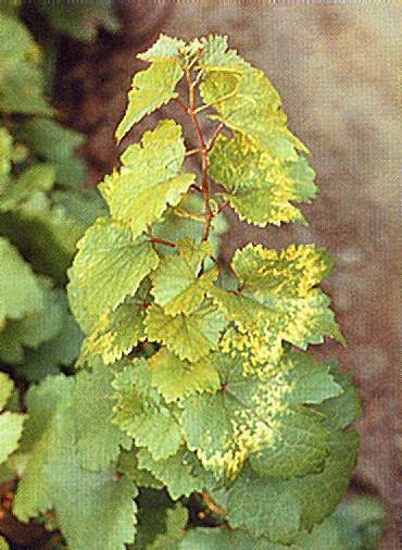
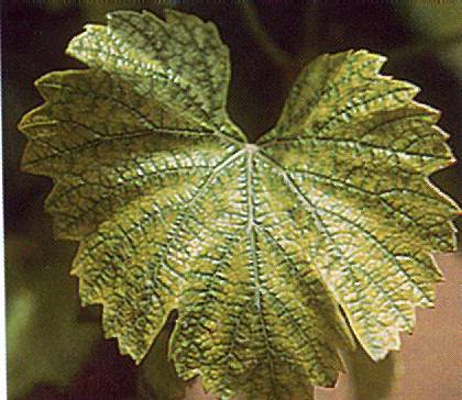
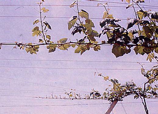
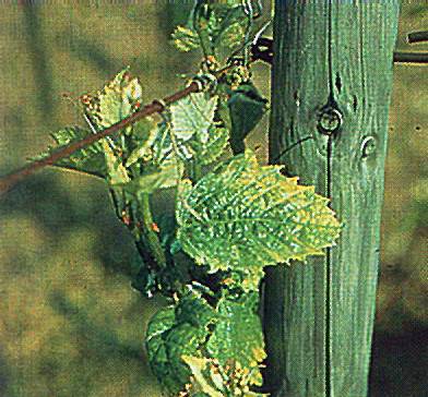
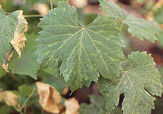
AMV infection
Bright yellow mottling induced by AMV infections in spring
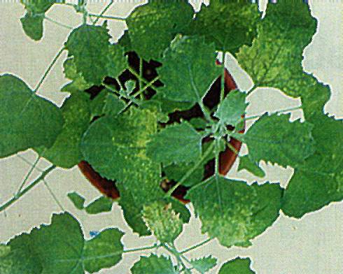
AMV_infection
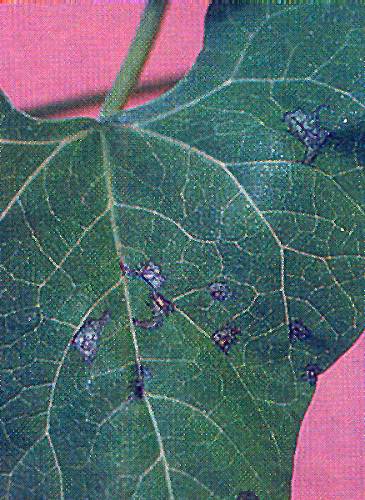
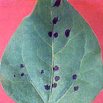
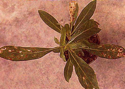
AMV infection
Necrotic local lesions induced by AMV in P. vulgaris (Photo: B. Walter)
AMV_infection
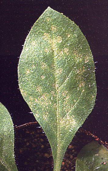
AMV infection
Necrotic local lesions induced by AMV in Vigna unguiculata (Photo: B. Walter)
AMV_infection
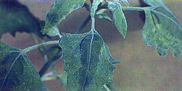
AMV infection
Systemic symptoms induced by AMV in C.quinoa (Photo: B. Walter)
AMV_Infection
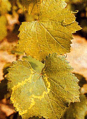
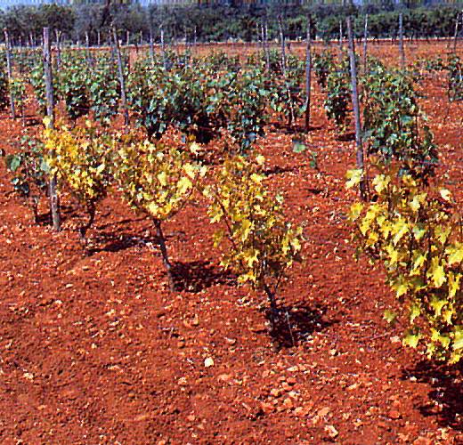
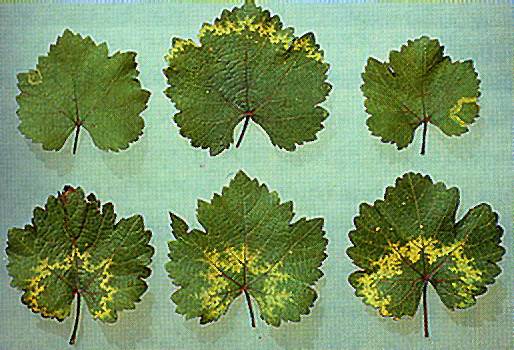
AMV_Infection
Yellow line pattern in graft-inoculated cv. Chardonnay indicator
AMV_infection
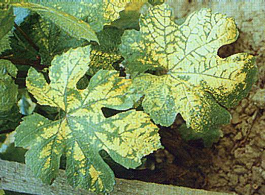
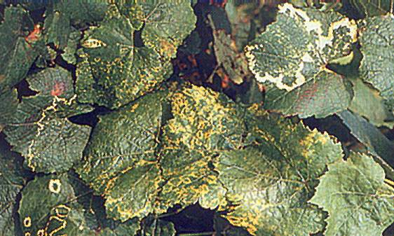
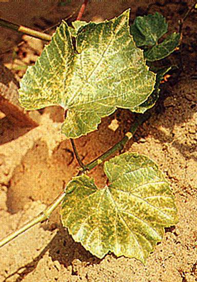
AMV infection
Yellow spots and line patterns in naturally infected cv. Chardonnay in summer
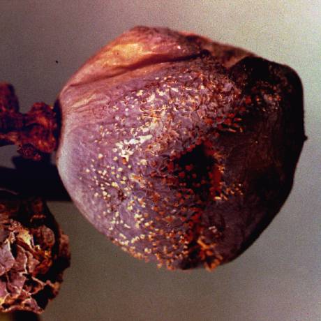
Anthracnose_Bird's_-_Eye_Rot
Anthracnose Bird's-Eye Rot
Anthracnose_Soft_Grape
Anthracnose Soft Grape
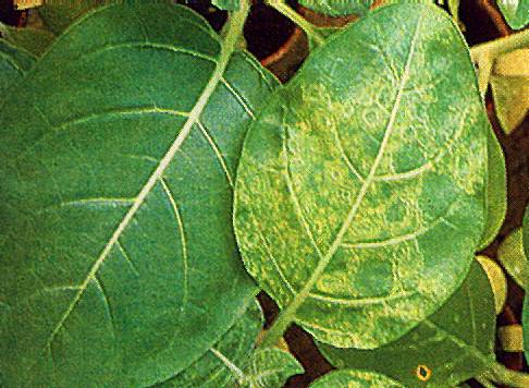
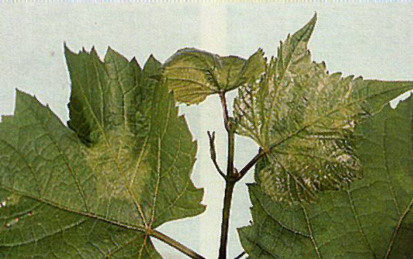
ArMV_induced_mosaic_mottle
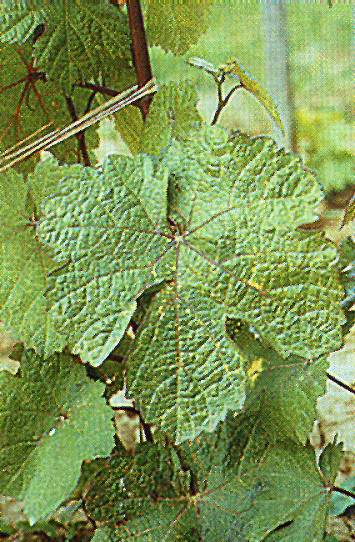
ArMV induced mosaic mottle
Mosaic mottle induced by ArMV in C.quinoa
ArMV_infection
ArMV infection
Mottling and deformation of a grape leaf induced by a distorting strain of ArMV
ArMV_infection
ArMV infection
Yellow mottling induced by a chromogenic strain of ArMV
ArMV_infection
ArMV infection
Yellow spots and rings induced by ArMV in N.glutinosa
Asteroid_mosaic
Asteroid mosaic
Foliar symptoms of asteroid mosaic (Photo: U.ota)
Asteroid_mosaic_virus_(AMV)_infection
Asteroid mosaic virus (AMV) infection
Foliar reactions of V. rupestris to asteroid mosaic infection
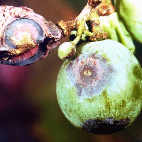
Bitter_Rot
Bitter Rot
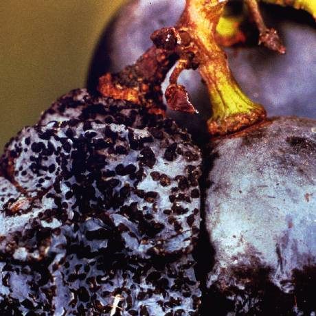
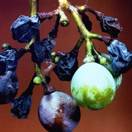
Black_Rot_on_Fruit
Black Rot on Fruit
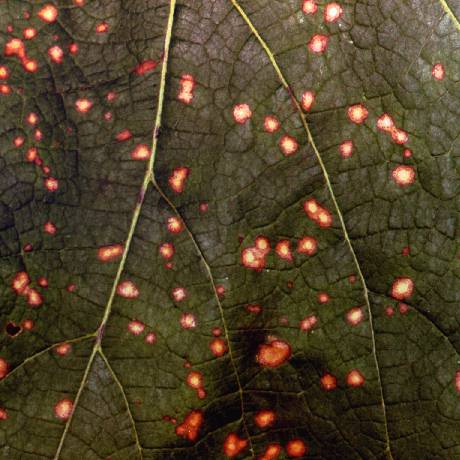
Black_Rot_on_Leaf
Black Rot on Leaf
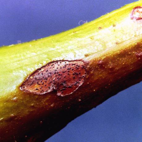
Black_Rot_on_Stem
Black Rot on Stem
Bois_noir_infection
Bois noir infection
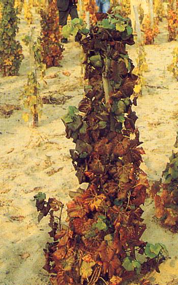
Leaf yellowing and drying up of bunches in a vine affected by bois noir (Photo: G. Granata)
Bois_noir_infection
Bois noir infection
Sectorial reddening typical of bois noir infection in a red-berried cultivar (Photo: G. Granata)
Bois_noir_infection
Bois noir infection
Severe yellowing of the leaf blade and necrosis of the main vein induced by bois noir infection in a white-berried cultivar (Photo: G. Granata)
Bois_noir_infection
Bois noir infection
View of a white-berried grapevine cultivar affected by bois noir (Photo: G. Granata)
CarMV_infection
CarMV infection
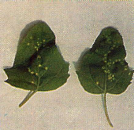
Local necrotic lesions and deformation of the upper leaves in G.globosa infected by the grapevine isolate of CarMV
CarMV_infection
CarMV infection
Necrotic spots and rings on a N.clevelandii leaf inoculated with the grapevine isolate of CarMV
Chrome_mosaic
Chrome mosaic
A row of vines with severe chrome mosaic symptoms
Chrome_mosaic_symptoms
Chrome mosaic symptoms
Chrome mosaic symptoms in the indicator Kober 5BB
Concord_grape
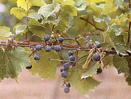
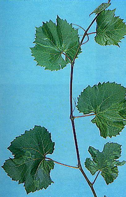
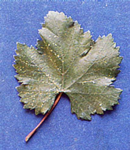
Concord grape
Healthy Concord vine
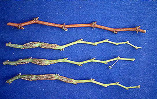
Corky_bark
Corky bark
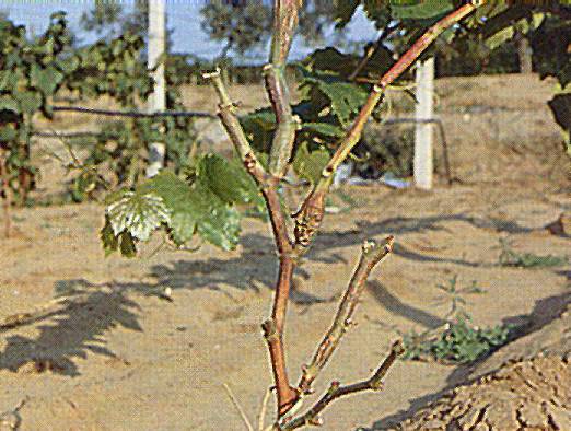
Canes from an LN33 indicator showing classical corky bark symptoms. A healthy mature cane (above) and immature canes with internodal swellings from graft-inoculated vine
Corky_bark
Corky bark
Symptoms shown by an LN 33 vine naturally infected following mealybug infestation in the field
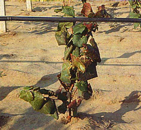
Corky_bark
Corky bark
Stunting and leaf reddening of an LN33 indicator affected by a severe form of corky bark
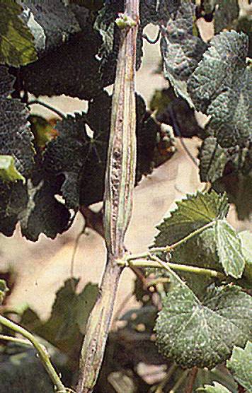
Corky_bark
Corky bark
Typical internodal swelling and cracking induced by corky bark in LN33
Corky_bark
Corky bark
Yellow spots sometimes appear in spring on leaves of LN33 graft-inoculated with corky bark sources
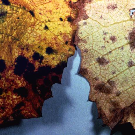
Cristulariella_1
Cristulariella 1
Cristulariella moricola, zonate leaf spot (John R. McGrew, USDA)
Downy_Mildew
Downy Mildew
Enations
Enations
Enations on indicator LN33 (Photo: U. Prota)
Enations
Enations
Enations on the underside of a leaf, clustered along the main veins
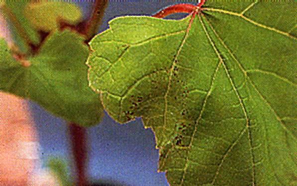
Enations
Enations
Severely malformed basal leaves of a European grape cultivar with outstanding enations
Fanleaf_infection
Fanleaf infection
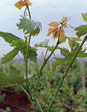
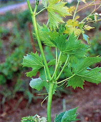
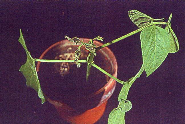
Abnormal branching in a shoot of a fanleaf-infected vine
Fanleaf_infection
Fanleaf infection
Abnormal shoot in a fanleaf-infected vine
Fanleaf_infection
Fanleaf infection
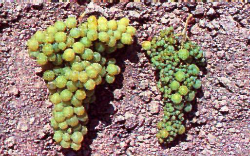
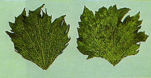
Bunches from a fanleaf-infected vine before (left) and after (right) heat therapy
Fanleaf_infection
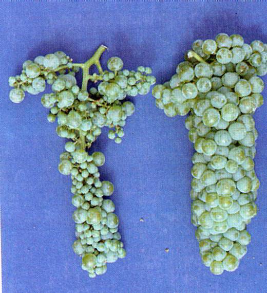
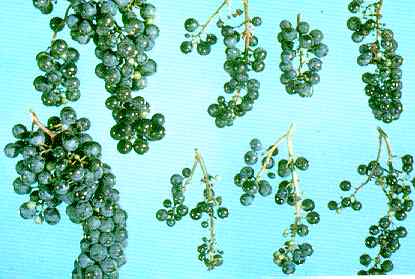
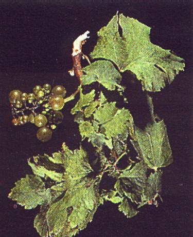
Fanleaf_infection
Bunches from a healthy (left) and a fanleaf-infected (right) vine. Note reduced size and extensive shot berry condition.
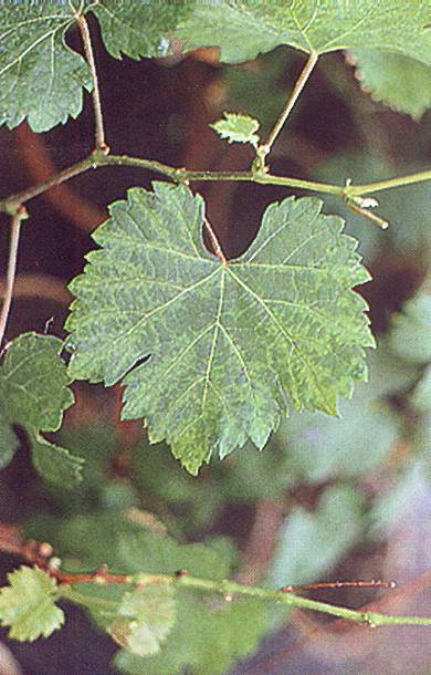
Fanleaf_infection
Fanleaf infection
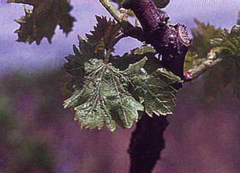
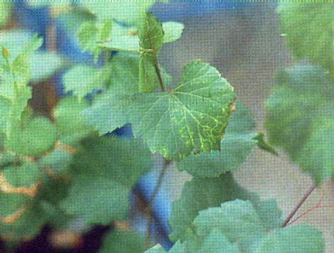
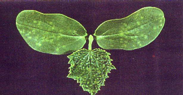
Chlorotic mottle and deformation caused by fanleaf infection
Fanleaf_infection
Fanleaf infection
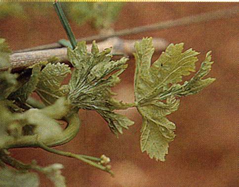
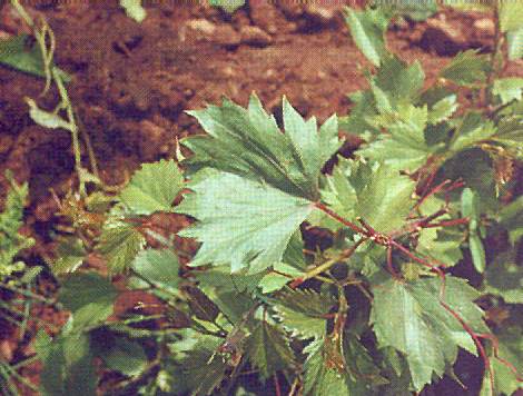
Chronic symptoms (severely deformed leaves with prominent teetn) in a V. rupestris indicator graft-inoculated with a fanleaf source
Fanleaf_infection
Fanleaf infection
Shock symptoms (chlorotic rings and lines) in a V.rupestris indicator graft-inoculated with a fanleaf source
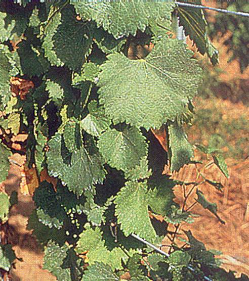
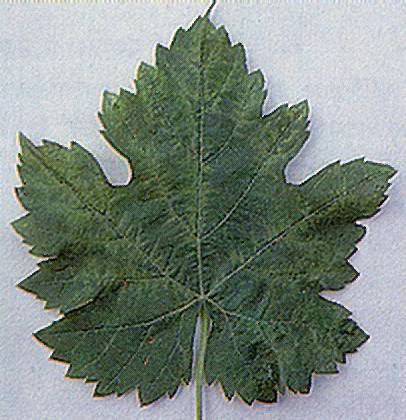
Fanleaf_symptoms
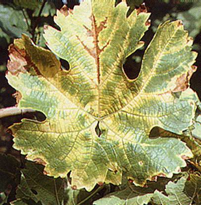
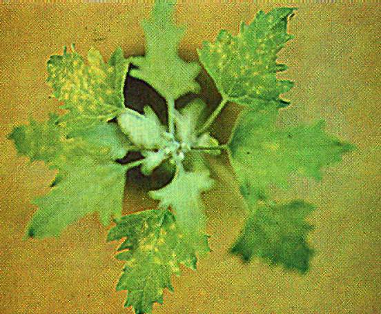
Fanleaf symptoms
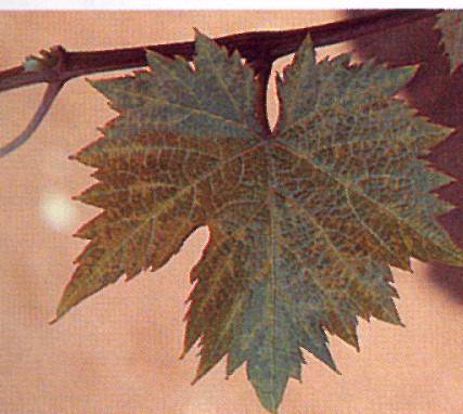
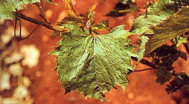
Grape leaf with typical fanleaf symptoms
Flavescence_doree_infection
Flavescence doree infection
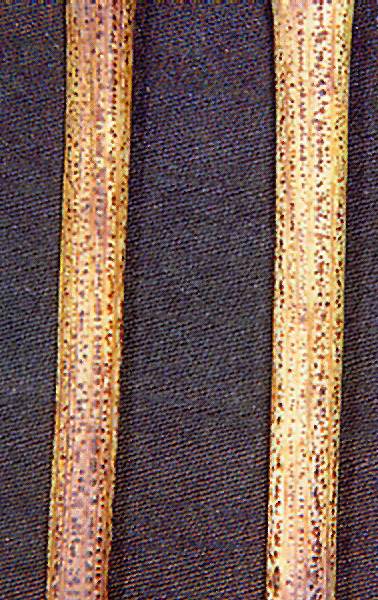
Black pustules on the canes of a vine infected by flavescence doree (Photo: G Granata)
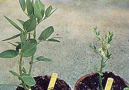
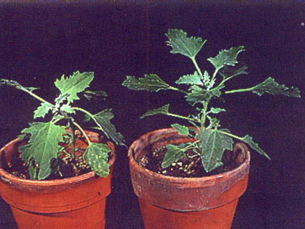
Flavescence_doree_infection
Flavescence doree infection
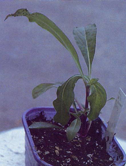
Broad bean plant artificially infected with the agent of flavescence doree (right) next to a healthy plant (left)
Flavescence_doree_infection
Flavescence doree infection
Close-up of Baco 22A leaves with symptoms of flavescence doree. Note shelling and shrivelling of the cluster
Flavescence_doree_infection
Flavescence doree infection
Instar of the flavescence doree vector Scaphoideus titanus (Photo: A. Brun)
Flavescence_doree_infection
Flavescence doree infection
Intense reddening caused by flavescence doree infection in cv. Pinot noir
Flavescence_doree_infection
Flavescence doree infection
Irregular wood ripening in a shot of a vine affected by flavescence doree
Flavescence_doree_infection
Flavescence doree infection
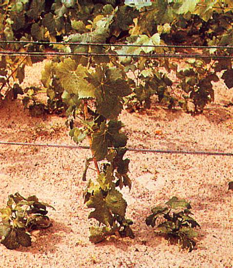
Reduced vigour, stunting and leaf reddening of cv. Aramon caused by flavescence doree
Flavescence_doree_infection
Flavescence doree infection
Vine of Baco 22A with stuntint and yellowing caused by flavescence doree infection (left) next to a healthy vine (right) (Photo: G. Granata)
Fleck_disease
Fleck disease
Clearing of the veinlets of V.rupestris typical of fleck disease
GCMV_infection
GCMV infection
Chlorotic local lesions induced by GCMV in C.quinoa
GCMV_infection
GCMV infection
Chlorotic mottling and leaf deformation induced by a distorting strain of GCMV
GCMV_infection
GCMV infection
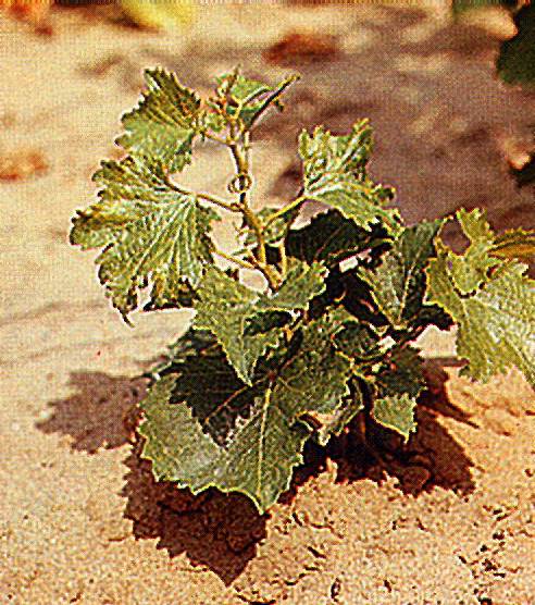
Extremely severe stunting caused by GCMV infections in the indicator Pinot noir two years after grafting
GCMV_infection
GCMV infection
Severe systemic yellowing induced by GCMV in C.quinoa
GCMV_infection
GCMV infection
Stunting, mottling and leaf deformation in a Siegfriedrebe indicator graft-inoculated with GCMV
GCMV_infection
GCMV infection
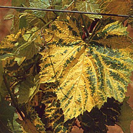
Typical yellow discoloration caused by a chromogenic strain of GCMV
GFLV_infection
GFLV infection
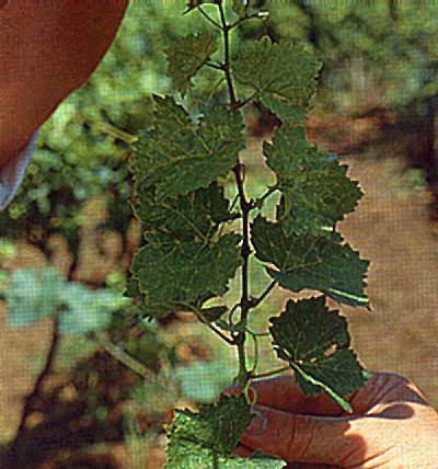
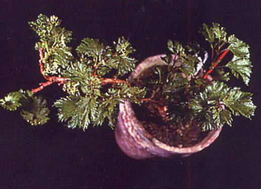
Severely malformed leaves and bushy vegetation in a vine infected by a distorting GFLV strain

GFLV_infection
GFLV_infection
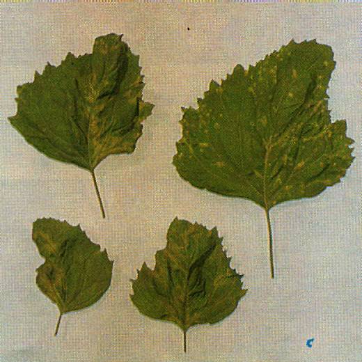
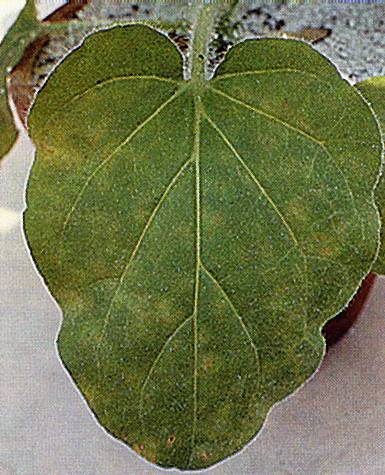
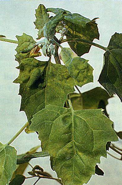
Systemic mottling and deformation of the leaves of Chenopodium quinoa infected by GFLV
GFLV_infection
GFLV infection
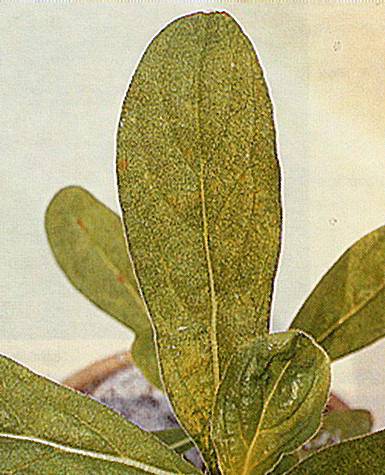
Systemic mottling induced by GFLV in Chenopodium amaranticolor
GFLV_infection
GFLV infection
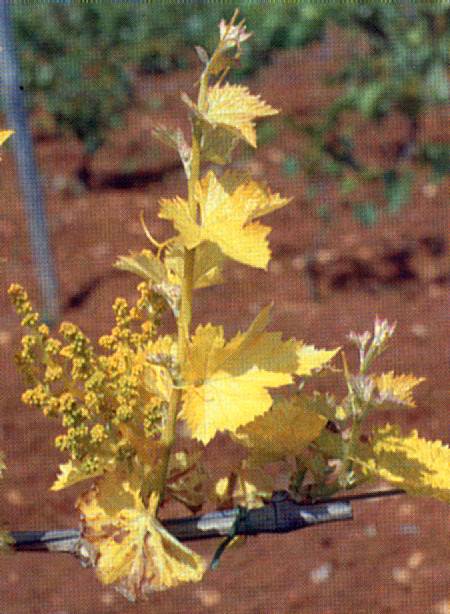
Total yellowing of a spring shoot of a vine infected by a chromogenic GFLV strain
GFLV_infection
GFLV infection
Twisting of the upper leaves typically induced by GFLV infections in Gomphrena globosa
GFLV_infection
GFLV infection
Yellow rings and line patterns in a leaf infected by a chromogenic GFLV strain
GLPV_infection
GLPV infection
Chlorotic local lesions in a N. glutinosa leaf inoculated with GLPV
GLPV_infection
GLPV infection
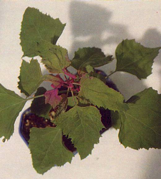
Distortion, mosaic and apical necrosis induced by GLPV in C.quinoa
GLPV_infection
GLPV infection
Young chlorotic lesions induced by GLPV in G.globosa
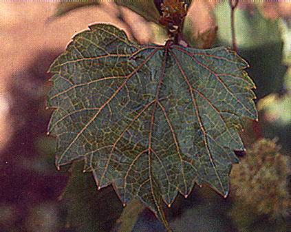
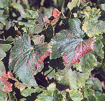
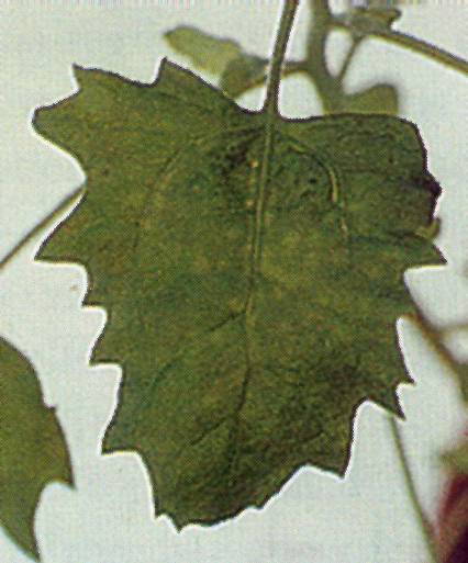
Grapevine_leafroll-associated
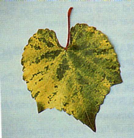
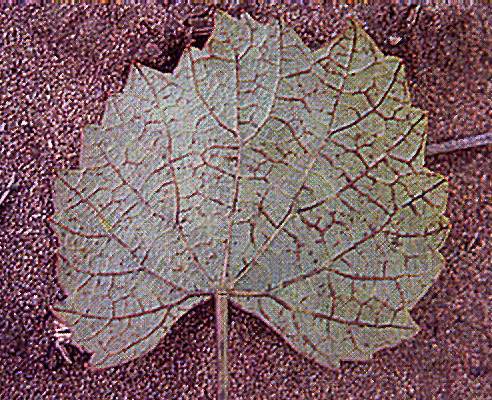
Grapevine leafroll-associated
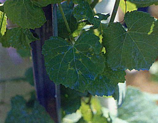
Grapevine leafroll-associated closteroviruses:
Interveinal chlorosis on white grape variety.

Grapevine_stunt
Grapevine stunt
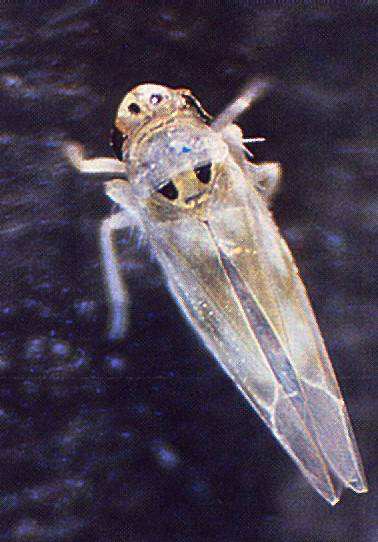
Adult of Arboridia apicalis, the leafhopper vector of grapevine stunt
GVA_infection
GVA infection
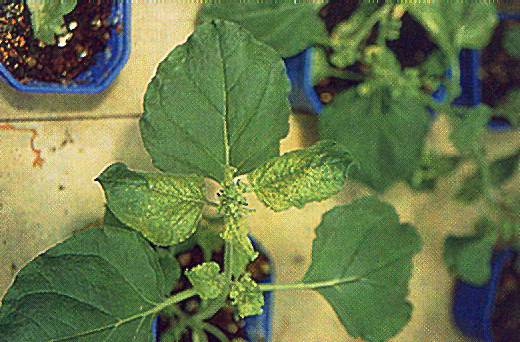
Vein yellowing in Nicotiana benthamiana infected with GVA
Isariopsis_Leaf_Spot
Isariopsis Leaf Spot
Leaf_blotch_1
Leaf blotch 1
Leaf blotch (caused, briosia ampelophaga), rougeon leaf (John R. McGrew, USDA)
Leaf_discoloration
Leaf discoloration
Reddish and yellow discolorations of the veins and margins of cv.Roditis leaves affected by leaf discoloration
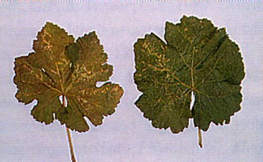
Leaf_discoloration
Leaf discoloration
Sectorial discolorations and vein yellowing in leaves of cv. Roditis vines affected by leaf discoloration
Leafroll_infection
Leafroll infection
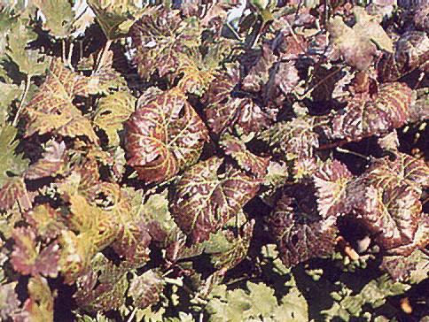
Discoloured leaves with main veins retaining the green colour
Leafroll_infection
Leafroll infection
Pale-berried bunches from a leafroll-infected vine
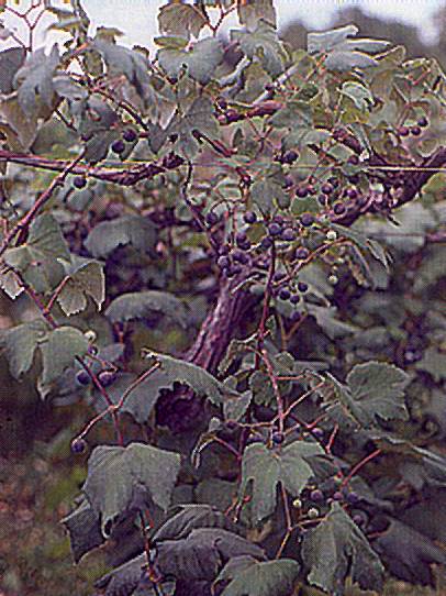
Leafroll_infection
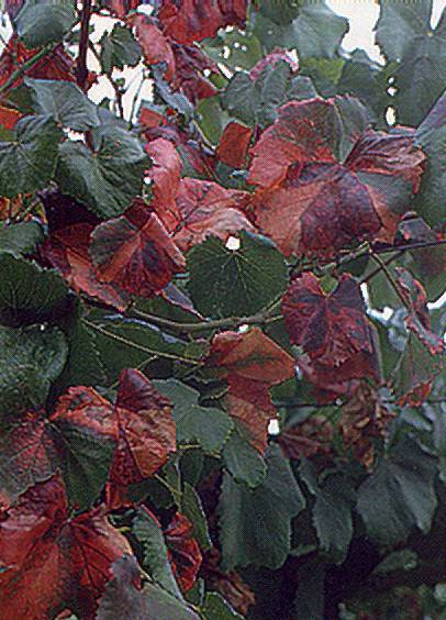
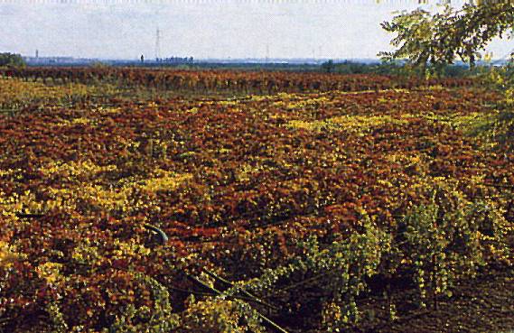
Leafroll infection
Red-fruited vines affected by leafroll are readily identified in the field in autumn
Leafroll_symptoms
Leafroll symptoms
Incipient rolling and reddening of the leaves in a vine infected by leafroll in spring
Leafroll_symptoms
Leafroll symptoms
Leafroll symptoms shown in autumn by an indicator vine two years after graft inoculation
Leafroll_symptoms
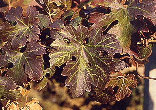
Leafroll symptoms
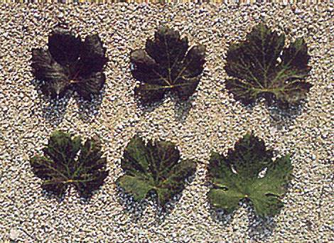
Progressive reddish discolorations in leaves from a vine affected by leafroll
Leafroll_symptoms
Leafroll symptoms
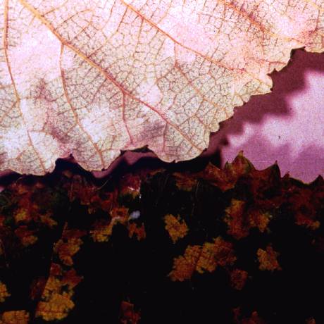
Severe leafroll symptoms shown in autumn by a red-fruited European grape cultivar.
Leafroll_symptoms
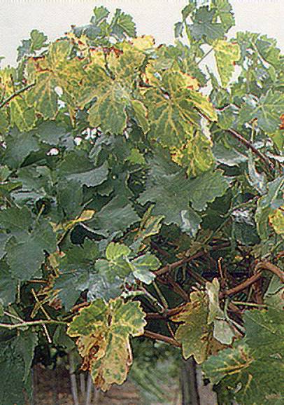
Leafroll symptoms
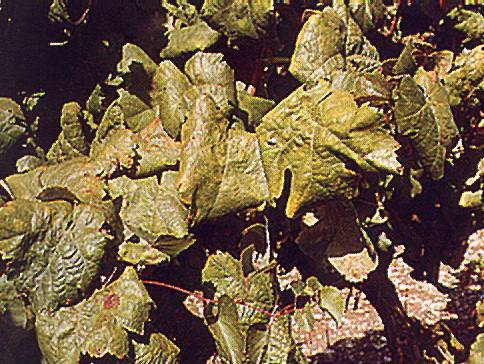
Yellowing and rolling of the leaves induced by leafroll in a white-fruited grape cultivar
Line_pattern_infection
Line pattern infection
Distribution of line pattern symptoms in a newly infected shoot of cv. Jubileum 75
Line_pattern_infection
Line pattern infection
Scattered minute yellow spots associated with chronic line pattern infection
Line_pattern_infection
Line pattern infection
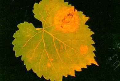
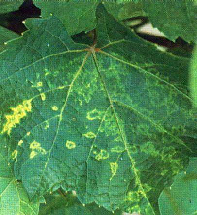
Yellow rings and lines typically associated with grapevine line pattern disease
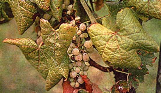
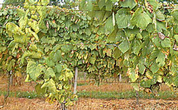
Pierce's_disease_(Xylella_fastidiosa)
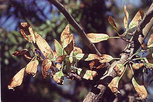
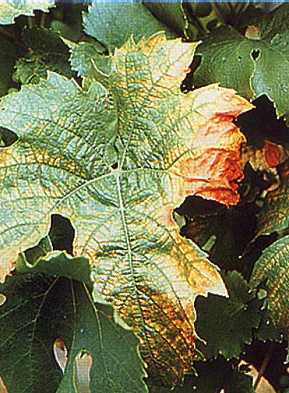
Pierce's disease (Xylella fastidiosa)
Chardonnay with Pierce's disease. A leaf in early spring exhibits characteristic interveinal chlorosis (Photo: P. Goodwin)
Pierce's_disease_(Xylella_fastidiosa)
Pierce's disease (Xylella_fastidiosa)
Close-up of autumn symptoms in cv. Chardonnay
Pierce's_disease_(Xylella_fastidiosa)
Pierce's disease (Xylella_fastidiosa)
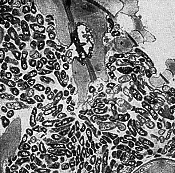
Electron micrograph of a cross-section through a xylem element of a Xylella fastidiosa-infected vine. The neighbouring vessel is free of bacteria (Photo: A. Purcell)
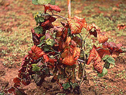
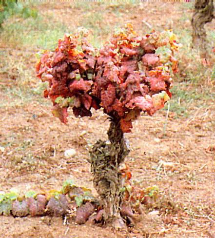
Pierce's_disease_(Xylella_fastidiosa)
Pierce's disease (Xylella_fastidiosa)
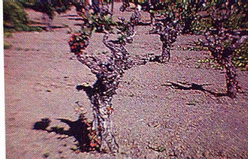
Final stage of a vine infected by Pierce's disease
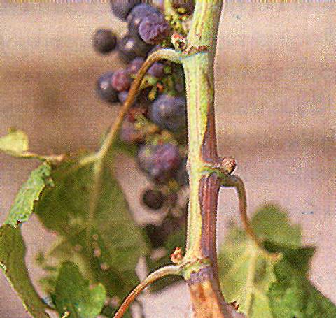
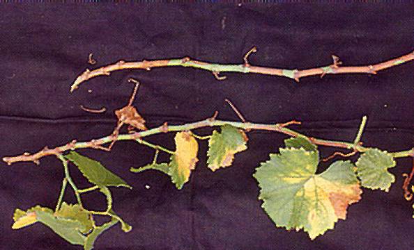
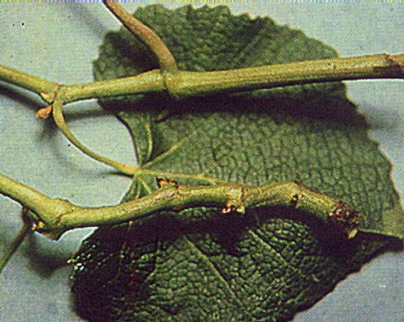
Pierce's_disease_(Xylella_fastidiosa)
Pierce's disease (Xylella fastidiosa)
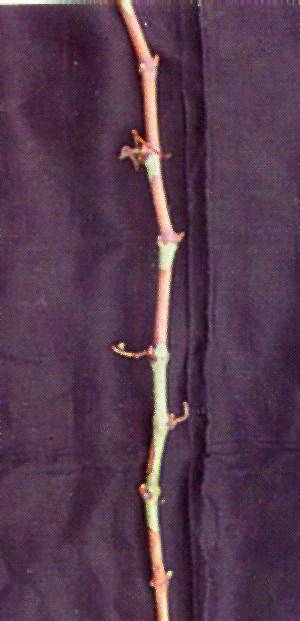
Green patches of immature wood alternating with brown mature wood in a cv. Chardonnay cane affected by Pierce's disease
Pierce's_disease_(Xylella_fastidiosa)
Pierce's disease (Xylella_fastidiosa)
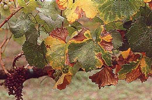
Marginal leaf scorching and uneven wood ripening of a cv. Chardonnay shoot affected by Pierce's disease
Pierce's_disease_(Xylella_fastidiosa)
Pierce's disease (Xylella fastidiosa)
Marginal scorching of cv. Merlot leaves caused by Pierce's dusease ub aytynb
Pierce's_disease_(Xylella_fastidiosa)
Pierce's disease (Xylella_fastidiosa)
Petioles persist after abscission of leaves on canes of a vine infected with Pierce's disease (Photo: H. Andris)

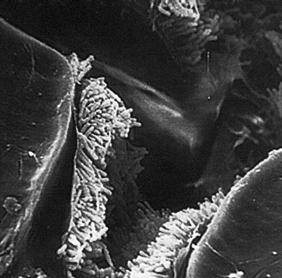
Pierce's_disease_(Xylella_fastidiosa)
Pierce's disease (Xylella fastidiosa)
Scanning electron micrograph of a mat of Xylella fastidiosa lining the aperture of the feeding stylet of a sharpshooter vector (Photo: M.G. Kinsey)
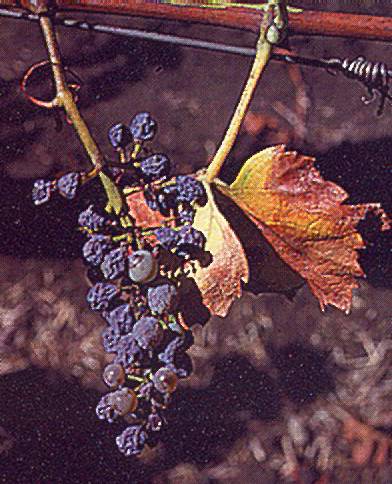
Pierce's_disease_(Xylella_fastidiosa)
Pierce's disease (Xylella_fastidiosa)
Shrivelling of a bunch in a vine affected by Pierce's disease. Note the green islands of bark resulting from uneven maturation of the canes (Photo: A. Yen)
Pierce's_disease_(Xylella_fastidiosa)
Pierce's disease (Xylella fastidiosa)
Symptoms of leaf scorching in almond infected with Xylella fastidiosa, the Pierce's disease agent
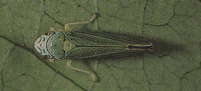
Pierce's_disease_(Xylella_fastidiosa)
Pierce's disease (Xylella fastidiosa)
The blue-green leafhopper Graphocephala atropunctata, one of the many sharpshooter species known to transmit Xylella fastidiosa (Photo: J. Clark)
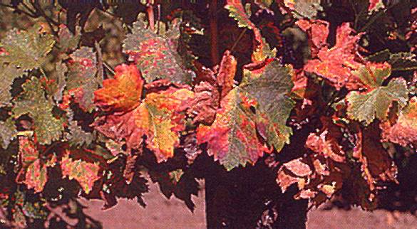
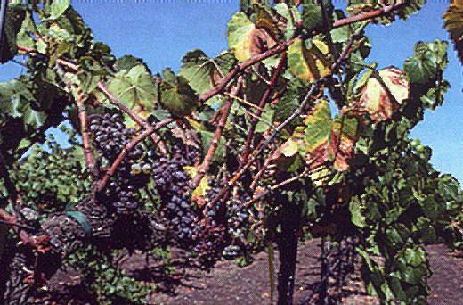
Pierce's_disease_(Xylella_fastidiosa)
Pierce's disease (Xylella fastidiosa)
Typical autumn symptoms of Pierce's disease in cv. Chardonnay in California (Photo: A. Yen)
PRMV_infection
PRMV infection
Irregular internodes and crooked canes consequent to PRMV infection in a Concord vine
PRMV_infection
PRMV infection
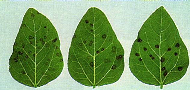
Necrotic local lesions induced by PRMV in C.quinoa
PRMV_infection
PRMV infection
Straggling clusters in a PRMV-infected vine
RRV_infection
RRV infection
Yellowing and marginal necrosis in vine infected by RRV
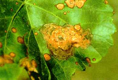
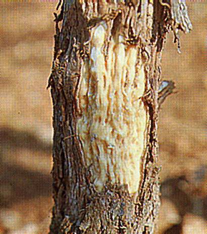
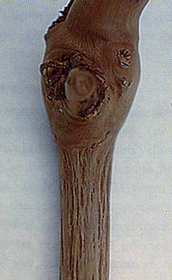
Rugose_wood
Rugose wood
A window open in the cortex at the level of the graft union to check the presence of rugose wood symptoms
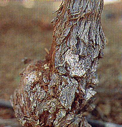
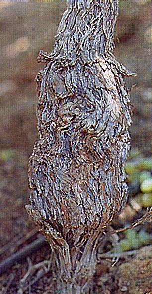
Rugose_wood
Rugose_wood
Corky appearance of the bark above the graft union in a vine affected by rugose wood
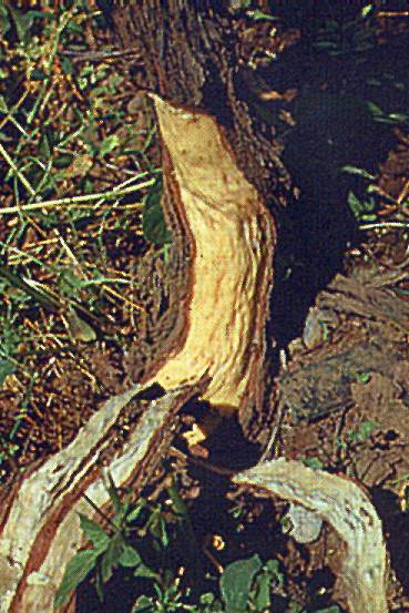
Rugose_wood
Rugose wood
Grooves can sometimes be seen on the outer surface of the cortex after removal of the bark
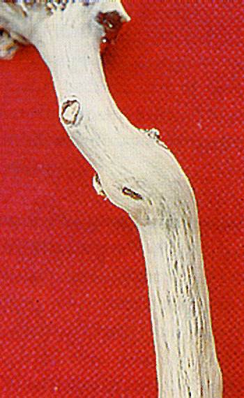
Rugose_wood
Rugose wood
Peg- and ridge-like protrusions on the cambial face of the peeled cortex of a diseased vine correspond to pits and grooves on the woody cylinder
Rugose_wood
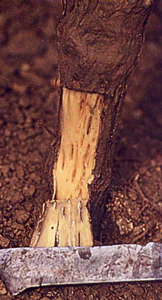
Rugose wood
Severe pitting of the trunk of a vine affected by rugose wood. The cortex has been removed to expose the altered woody cylinder
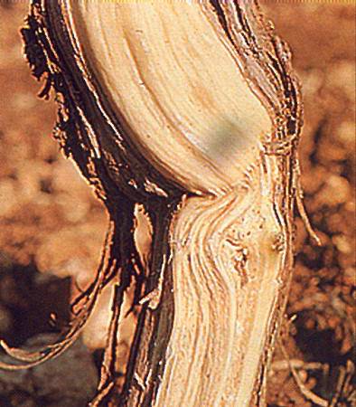
Rugose_wood
Rugose wood
Swelling above the graft union in a vine affected by rugose wood. Note the difference in diameter between scion and rootstock and the rough appearance of the bark
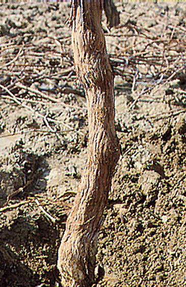
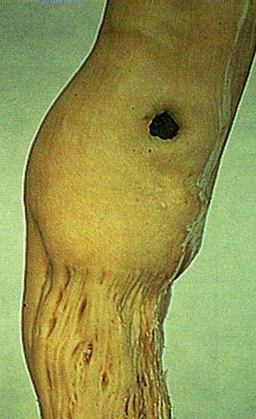
Rugose_wood
Rugose wood
Symptoms of rugose wood showing on both scion and rootstock of a diseased vine
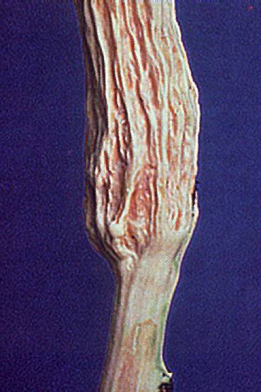
Rugose_wood
Rugose wood
Symptoms of rugose wood showing only on the rootstock of a diseased vine
Rugose_wood
Rugose wood
Symptoms of rugose wood showing only on the scion of a diseased vine
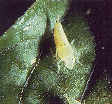
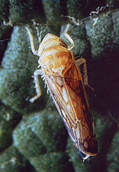
Scaphoideus_titanus
Scaphoideus titanus
Adult of Scaphoideus titanus (Photo: A. Brun)
SLRV_infection
SLRV infection
Chlorotic local lesions induced by SLRV in C.quinoa
SLRV_infection
SLRV infection
Reddish discolorations of the shoot apex typically induced by SLRV in spring in certain cultivars (left) compared to healthy shoot (right)
SLRV_infection
SLRV infection
Summer symptoms of SLRV. The vegetation is apparently normal but there is no crop
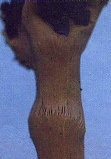
Stem_grooving
Stem grooving
Stem grooving in a Kober 5BB indicator. Note absence of symptoms on the scion
Stem_grooving
Stem grooving
Stem grooving in an LN 33 indicator that did not show secondary phloem proliferation. Note absence of symptoms on the scion
Stem_pitting_disease
Stem pitting disease
Typical strip of basipetal pitting extending downward from the point of inoculation in a V.rupestris indicator inoculated by chip-budding with a source of rupestris stem pitting disease
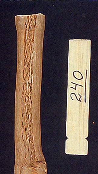
Stem_pitting_symptoms
Stem pitting symptoms
Rupestris stem pitting symptoms in a V.rupestris indicator inoculated by top grafting. The pits are all around and just below the graft union
Stunt_symptoms
Stunt symptoms
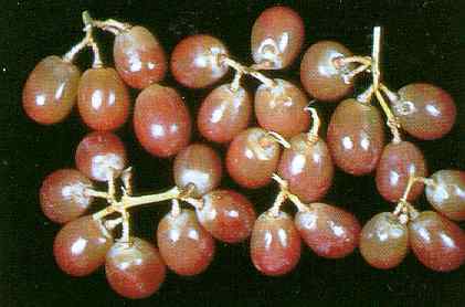
Bunches of cv. Koshu from a diseased (left) and a healthy (right) vine. Note the whitish colour and the smaller berries of the diseased bunch
Stunt_symptoms
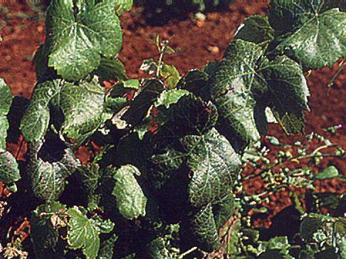
Stunt symptoms
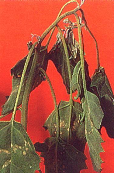
Stunt symptoms in spring. Note poor growth and curling of the leaves
Sulfur_dioxide_1
Sulfur dioxide 1
Sulfur dioxide damage on emperor grapes, bleached, sunken tissue (J. Harvey, USDA)
TBRV_infection
TBRV infection
Necrotic local lesions and top necrosis induced by TBRV in C.quinoa
TBVR_infection
TBVR infection
Leaf deformity and yellowing in a vine infected by TBRV
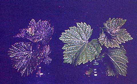
Tomato_ringspot_2
Tomato ringspot 2
Tomato ringspot virus (right) on cascade berry clusters, healthy vines (left) (J. K. Uyemoto, USDA)
ToRsv_infection
ToRsv infection
Chlorotic rings and leaf deformation in a Baco shoot infected by ToRSV
ToRSV_infection
ToRSV infection
Shock reaction of a Baco indicator to ToRSV infection: chlorotic blotch
ToRSV_infection
ToRSV infection
Shock reaction of a Kober 5BB indicator to ToRSV infection: chlorotic blotch with necrotizing margin
ToRSV_infection
ToRSV infection
ToRSV-infected Concord vine. Note the markedly reduced crop
ToRSV_infection
ToRSV infection
Typical yellow vein symptoms elicited by the California strain of ToRSV
TRSV_and_GFLV_infections
TRSV and GFLV infections
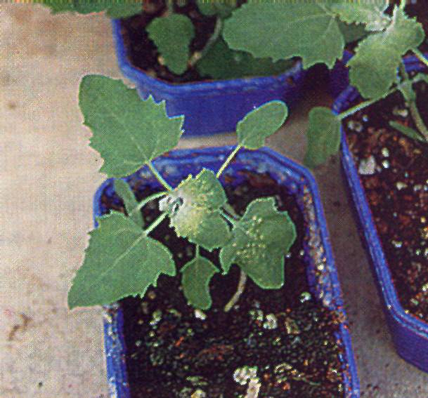
Plants of Chenopodium quinoa 9 days after inoculation with TRSV (left) and GFLV (right)
TRSV_infection
TRSV infection
Chenopodium amaranticolor leaf with chlorotic lesions 9 days after inoculation with TRSV (left)

TRSV_infection
TRSV infection
Cucumber plant a week after inoculation with TRSV showing chlorotic lesions on the cotyledons and systemic mottle
TRSV_infection
TRSV infection
Extremely severe symptoms in a Chardonnay indicator inoculated with a TRSV source
TRSV_infection
TRSV infection
Leaves from a declining Chardonnay vine infected by TRSV. These symptoms are indistinguishable from those induced by GFLV and other European nepoviruses
TRSV_infection
TRSV infection
Necrotic lesions on leaves of cowpea inoculated with TRSV
TRSV_infection
TRSV infection
Phaseolus vulgaris with tip necrosis two weeks after inoculation with TRSV
TRSV_infection
TRSV infection
Shoot of a field-grown Chardonnay vine infected by TRSV
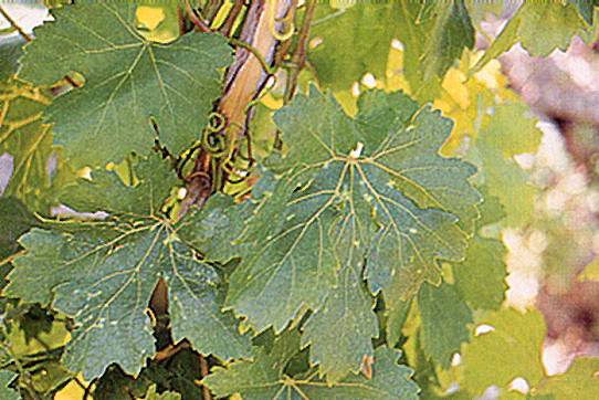
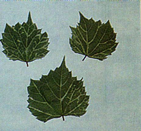
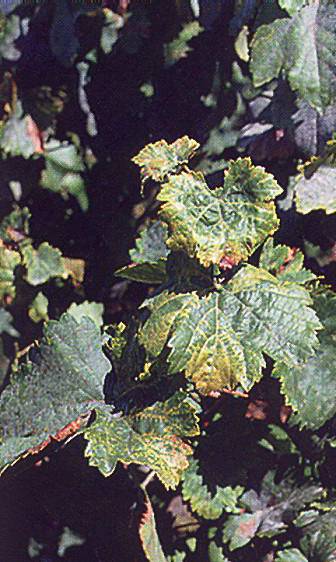
Vein_mosaic_disease
Vein_mosaic_disease
Chlorotic vein feathering induced by vein mosaic disease
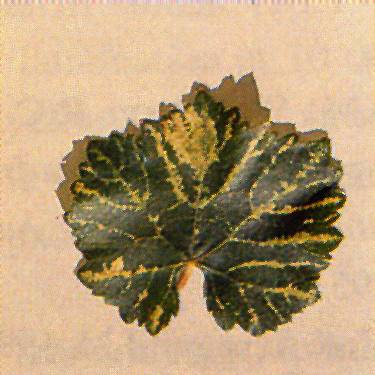
Vein_mosaic_disease
Vein mosaic disease
Intense chlorotic vein banding induced by vein mosaic disease
Vein_mosaic_disease
Vein mosaic disease
Vein mosaic symptoms in the indicator V. riparia
Vein_necrosis
Vein necrosis
Necrosis of the veinlets typically induced by vein necrosis in the indicator 110 R
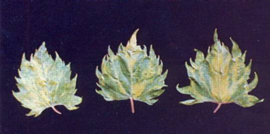
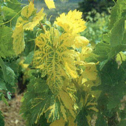
Yellow_mosaic
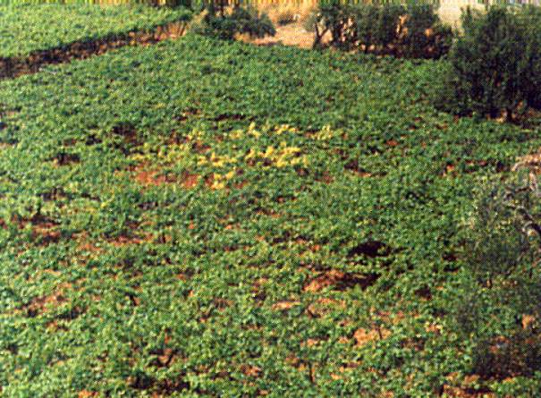
Yellow mosaic
A patch of vines with yellow mosaic seen from a distance
Yellow_mosaic
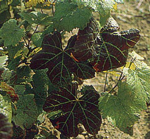
Yellow mosaic
Chrome yellow discolorations and deformations of leaves of a V. rupestris indicator graft-inoculated with a yellow mosaic source
Yellow_mosaic
Yellow mosaic
Spring symptoms of yellow mosaic
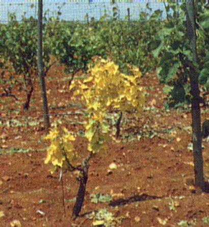
Yellow_mosaic
Yellow mosaic
Totally yellow and stunted vine affected by yellow mosaic next to a normally growing plant
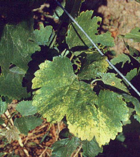
Yellow_mosaic
Yellow mosaic
Yellow mosaic symptoms fading away in late summer
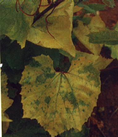
Yellow_mosaic
Yellow mosaic
Yellow mosaic symptoms in a leaf of an American rootstock
Yellow_speckle_and_leafroll
Yellow speckle and leafroll
Symptoms of yellow speckle and leafroll appearing in autumn on the same vine
Yellow_speckle_infection
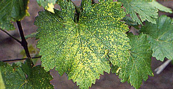
Yellow speckle infection
Scattered yellow spots in a European grape leaf, typical of yellow speckle infection
Yellow_speckle_infection
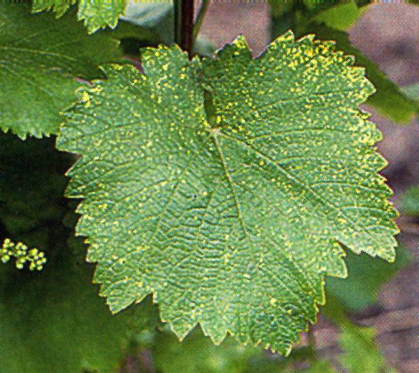
Yellow speckle infection
Strong yellow speckle symptoms with speckles tending to gather along the main veins
Yellow_speckle_infection
Yellow speckle infection
Symptoms as in Figure 104, but with more intense yellow speckling
Yellow_speckle_viroid_and_grapevine_fanleaf_virus
Yellow speckle viroid and grapevine fanleaf virus
Vein banding symptoms in a vine doubly infected with yellow speckle viroid and grapevine fanleaf virus
Yellow_speckle_viroid_and_grapevine_fanleaf_virus
Yellow speckle viroid and grapevine fanleaf virus
Vein banding symptoms in a vine doubly infected with yellow speckle viroid and grapevine chrome mosaic virus (Photo: J. Lehoczky)