Pit Fruits:Photos
Pit/Stone Fruit Pest and Nutritional Disorder Photos
Contents
- 1 Alternaria_Fruit_Rot_1
- 2 Alternaria_Fruit_Rot_2
- 3 American_Brown_Rot_1
- 4 American_Brown_Rot_2
- 5 American_Brown_Rot_3
- 6 American_Brown_Rot_4
- 7 American_Brown_Rot_5
- 8 Anthracnose_1a Anthracnose 1a
- 9 Anthracnose_1b
- 10 Apricot,_2,4-D_herbicide_injury
- 11 Apricot,_Alternaria_spot,_fruit_symptoms
- 12 Apricot,_asteroid_spot,_leaf_symptoms
- 13 Apricot,_Coryneum_fruit_infection
- 14 Apricot,_hail_damage,_fruit
- 15 Apricot,_powdery_mildew,_russet
- 16 Apricot,_Roundup_herbicide_damage
- 17 Apricot_decline,_on_plum_rootstock_(incompatibility)
- 18 Apricot_ring_pox_virus_on_fruit
- 19 Apricot_ring_pox_virus_on_fruit_
- 20 Apricot_ring_pox_virus_on_fruit_(lesions)
- 21 Apricot_ring_pox_virus_on_leaves_(red_blotched_veins)
- 22 Armillaria_and_Clitocybe_Root_Rots_1
- 23 Armillaria_and_Clitocybe_Root_Rots_2a
- 24 Armillaria_and_Clitocybe_Root_Rots_2b
- 25 Armillaria_and_Clitocybe_Root_Rots_3
- 26 Armillaria_and_Clitocybe_Root_Rots_4
- 27 Bacteral_Spot_of_Leaf
- 28 Bacterial_Canker_1a
- 29 Bacterial_Canker_1b
- 30 Bacterial_Canker_2
- 31 Bacterial_Canker_3
- 32 Bacterial_Canker_4
- 33 Bacterial_Canker_5
- 34 Bacterial_Canker_6
- 35 Bacterial_Spot_1
- 36 Bacterial_Spot_2
- 37 Bacterial_Spot_3
- 38 Bacterial_Spot_4a
- 39 Bacterial_Spot_4b
- 40 Bacterial_Spot_5
- 41 Bacterial_Spot_6
- 42 Bacterial_Spot_on_Fruit
- 43 Bing_cherry,_X-disease,_graft_union_symptoms_(Mahaleb_rootstock)
- 44 Black_Knot_of_Plum_-_Plum
- 45 Black_Knot_of_Plum_1a
- 46 Black_Knot_of_Plum_1b
- 47 Black_Knot_of_Plum_2
- 48 Blast_Disease_on_Trunk
- 49 Blossom_Blight
- 50 Brown_Rot_on_Fruit
- 51 Cherry,_Coryneum_leaf_infection
- 52 Cherry,_2,4-D_herbicide_injury
- 53 Cherry_Leaf_Spot_1
- 54 Cherry_Leaf_Spot_2a
- 55 Cherry_Leaf_Spot_2b
- 56 Cherry_Leaf_Spot_3
- 57 Cherry_Leaf_Spot_4
- 58 Cherry_Leaf_Spot_5
- 59 Cherry_Leaf_Spot_6
- 60 Cherry_Leaf_Spot_7a
- 61 Cherry_Leaf_Spot_7b
- 62 Cherry_Leaf_Spot_8
- 63 Cherry_mottle_leaf_virus
- 64 Cherry_mottle_leaf_virus_on_sweet_cherry
- 65 Cherry_rusty_mottle_disease
- 66 Cherry_stem_pitting_(virus),_pitting_symptoms_at_graft_union
- 67 Cherry_stem_pitting(virus),_whole_tree_collapse
- 68 Cherry_tree,_weak
- 69 Cherry_twisted_leaf_virus,_leaf_symptoms
- 70 Cherry_X-disease_(little_cherry),_symptomatic_vs_normal
- 71 Cherry_X-disease_(little_cherry)_fruit_symptoms_on_Lambert
- 72 Cherry_X-disease_(little_cherry)_leaf_symptoms
- 73 Chokecherry,_1st_year_symptoms_on_leaves
- 74 Chokecherry,_X-disease,_early_leaf_coloration_symptoms
- 75 Chokecherry,_X-disease,_early_leaf_coloration_symptoms
- 76 Chokecherry,_X-disease,_symptoms
- 77 Crinkle_leaf
- 78 Crinkle_leaf
- 79 Crown_Gall_1
- 80 Crown_Gall_2a
- 81 Crown_Gall_2b Crown Gall 2b
- 82 Cycle_brown_rot
- 83 Cycle_cherry_leaf_spot
- 84 Cycle_peach_bacterial_spot
- 85 Deep_suture_symptoms
- 86 Early_Red_Haven_peach,_peach_mosaic_rosetting_on_right
- 87 Elberta_peach,_peach_wart,_fruit_symptoms
- 88 Elberta_peach,_peach_wart,_fruit_symptoms
- 89 European_Brown_Rot_1
- 90 European_Brown_Rot_2
- 91 European_Brown_Rot_3
- 92 Fusicoccum_Canker_1
- 93 Fusicoccum_Canker_2
- 94 Gibertella_Rot
- 95 Graft_union_incompatibility_(TmRS)
- 96 Graft_union_of_apricot_decline_on_plum_rootstock
- 97 Green_Ring_Mottle_1
- 98 Green_Ring_Mottle_2
- 99 Gummosis
- 100 Lambert_sweet_cherry,_little_cherry_virus,_fruit_symptoms
- 101 Loring_peach,_A._healthy,_B._symptomatic
- 102 Montmorency_sour_cherry_+_sap_exudation,_ethrel_treated_@_2000_ppm
- 103 Morello_sour_cherry,_crown_borer_injury
- 104 Morello_sour_cherry,_crown_borer_injury_on_right
- 105 Orchard_Disease_Problem
- 106 Peach,_Botryosphaeria_canker,_bark_tissue_symptoms
- 107 Peach,_Botryosphaeria_canker,_killed_tree_with_severe_gumming
- 108 Peach,_brown_rot_mummy
- 109 Peach,_burr_knot_(aerial_crown_gall)
- 110 Peach,_chronic_chlorosis_(iron_deficiency),_tree_dieback
- 111 Peach,_Complete_Green,_phytotoxicity
- 112 Peach,_Complete_Green,_phytotoxicity,_leaf_injury
- 113 Peach,_Coryneum_blight,_fruit_infection
- 114 Peach,_Coryneum_blight,_fruit_infection
- 115 Peach,_Coryneum_blight,_fruit_infection
- 116 Peach,_Coryneum_blight,_fruit_infection_(late_season)
- 117 Peach,_Cytospora_canker,_infection_process
- 118 Peach,_Cytospora_canker_(gummosis)_symptoms
- 119 Peach,_Cytospora_canker_(gummosis)_symptoms
- 120 Peach,_Cytospora_canker_+_sunscald
- 121 Peach,_Cytospora_canker_infection_at_pruning_cuts
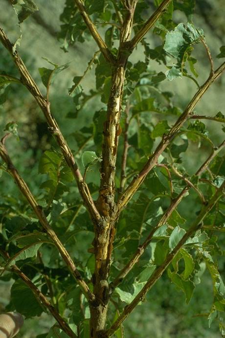
- 122 Peach,_freeze_&_winter_injury,_shoots
- 123 Peach,_hail_damage
- 124 Peach,_hail_damage,_fruit
- 125 Peach,_hail_damage,_tree_bark
- 126 Peach,_hail_damage,_tree_bark
- 127 Peach,_herbicide_damage
- 128 Peach,_herbicide_damage
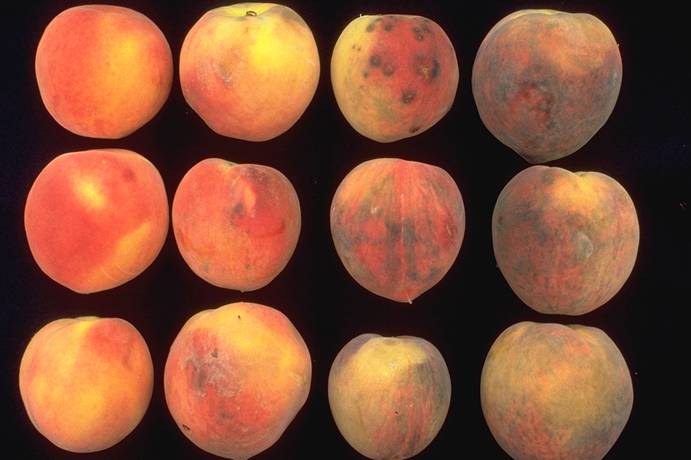
- 129 Peach,_inking_(abiotic)_-_severe_on_right
- 130 Peach,_iron_deficiency_chlorosis_control_with_iron_chelate
- 131 Peach,_Kearneyville_free_Y,_perpendicular_V
- 132 Peach,_leaf_cover_+_herbicide_timing
- 133 Peach,_left_dead,_right_viable_&_unopened
- 134 Peach,_peach_twig_borer_damage
- 135 Peach,_powdery_mildew,_fruit_infection_(rusty_spot)_
- 136 Peach,_range_of_foliage_colors,_winter_injury_recovery_+_fertilizer_trts
- 137 Peach,_red_suture_(MLO),_fruit_symptoms
- 138 Peach,_red_suture_(MLO),_leaf_&_fruit_symptoms
- 139 Peach,_Rhizopus,_5_days_post-harvest
- 140 Peach,_Rhizopus_rot_of_fruit
- 141 Peach,_severe_chlorosis_(iron_deficiency),_tree_dieback
- 142 Peach,_shot-hole_borer_+_woodpecker_feeding
- 143 Peach,_snow_damage
- 144 Peach,_snow_damage
- 145 Peach,_trunk_cracking/spiral
- 146 Peach,_western_X_disease,_whole_tree_symptoms
- 147 Peach,_winter_bud_kill
- 148 Peach,_winter_injury,_wood_browning
- 149 Peach,_winter_injury_+_pruning_timing_effects
- 150 Peach,_winter_injury_on_left_(pruned)
- 151 Peach,_X-disease_+_winter_damage
- 152 Peach,_X-disease_+_winter_damage
- 153 Peach,_X-disease_+_winter_damage
- 154 Peach,_X-disease_+_winter_damage
- 155 Peach_Leaf_Curl_1
- 156 Peach_Leaf_Curl_2
- 157 Peach_Leaf_Curl_3a
- 158 Peach_Leaf_Curl_3b
- 159 Peach_Leaf_Curl_4a
- 160 Peach_Leaf_Curl_4b
- 161 Peach_Leaf_Curl_4c
- 162 Peach_mosaic,_flower_color_break_symptom
- 163 Peach_mosaic,_flower_color_break_symptom
- 164 Peach_mosaic,_foliage_symptoms
- 165 Peach_mosaic,_foliage_symptoms_(isolate_CL-1)
- 166 Peach_mosaic,_foliage_symptoms_(isolate_CL-2)
- 167 Peach_mosaic,_Loring_peach,_L._healthy,_R._symptomatic_
- 168 Peach_mosaic,_peach_&_nectarine_fruit_symptoms
- 169 Peach_mosaic,_vein_clearing_symptoms
- 170 Peach_mosaic_leaf_symptoms,_healthy_vs_infected
- 171 Peach_Perennial_Canker_1
- 172 Peach_Perennial_Canker_2
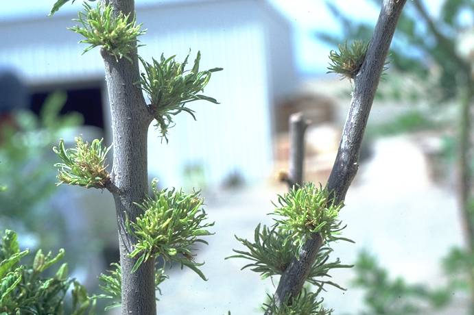
- 173 Peach_rosette_MLO
- 174 Peach_Rosette_Mosaic
- 175 Peach_Scab_1
- 176 Peach_Scab_2
- 177 Peach_Scab_3
- 178 Peach_winter_damage,_pruned_before_2/6/89
- 179 Peach_yellow_bud_strain_of_tomato_ringspot_virus
- 180 Peach_yellows_MLO
- 181 Phytophthora_Root,_Crown,_and_Colar_Rot_1
- 182 Phytophthora_Root,_Crown,_and_Colar_Rot_2
- 183 Plum_Pockets_1
- 184 Plum_Pockets_2
- 185 Plum_pox_virus
- 186 Powdery_Mildew/Rusty_Spot_of_Peach_1
- 187 Powdery_Mildew/Rusty_Spot_of_Peach_2a
- 188 Powdery_Mildew/Rusty_Spot_of_Peach_2b
- 189 Powdery_Mildew_of_Cherry_1
- 190 Powdery_Mildew_of_Cherry_2
- 191 Powdery_Mildew_of_Cherry_3
- 192 Powdery_mildew_spraying_(handgun_application)
- 193 Prune_dwarf_virus_symptoms_on_Italian_prune
- 194 Prune_dwarf_virus_symptoms_on_Italian_prune
- 195 Prunus_necrotic_ringspot_ilarvirus
- 196 Prunus_Necrotic_Ringspot_1
- 197 Prunus_Necrotic_Ringspot_2
- 198 Prunus_Stem_Pitting/Prune_Brownline_1
- 199 Prunus_Stem_Pitting/Prune_Brownline_2
- 200 Prunus_Stem_Pitting/Prune_Brownline_3a
- 201 Prunus_Stem_Pitting/Prune_Brownline_3b
- 202 Prunus_Stem_Pitting/Prune_Brownline_4
- 203 Rhizopus
- 204 Root_Rot
- 205 Rusty_blotch
- 206 Scab_on_Fruit
- 207 Scab_on_Twig
- 208 Silver_Leaf
- 209 Sour_cherry,_crown_borer_damage
- 210 Sour_cherry,_powdery_mildew,_leaf_infection
- 211 Sour_cherry,_snow_damage
- 212 Sour_cherry_decline,_crown_borer
- 213 Sour_cherry_decline,_crown_borer
- 214 Sour_cherry_decline,_crown_borer_injury
- 215 Sour_Cherry_Yellows
- 216 Sweet_cherry,_cherry_rasp-leaf_virus,_symptoms
- 217 Sweet_cherry,_Coryneum_leaf_infection
- 218 Sweet_cherry,_Cytospora_collapse
- 219 Sweet_cherry,_Cytospora_fruiting_bodies_beneath_bark_epidermis
- 220 Sweet_cherry,_Cytospora_infection_progress
- 221 Sweet_cherry,_powdery_mildew_on_leaf_(healthy_left)
- 222 Sweet_cherry,_Prunus_rusty_mottle_virus,_leaf_symptoms
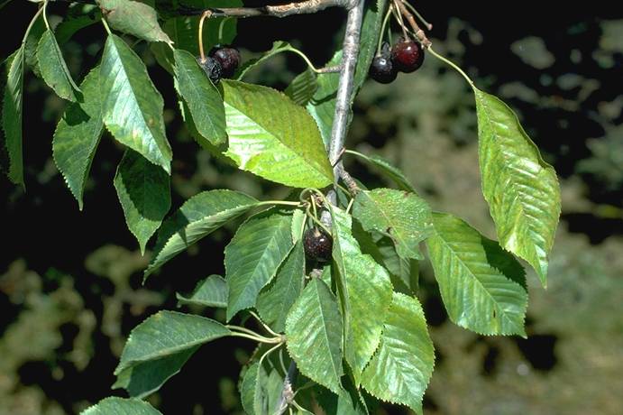
- 223 Sweet_cherry,_Prunus_rusty_mottle_virus,_tree_symptoms
- 224 Sweet_cherry,_Verticillium_wilt,_whole_tree_symptoms
- 225 Sweet_cherry,_X-disease,_fruit_symptoms
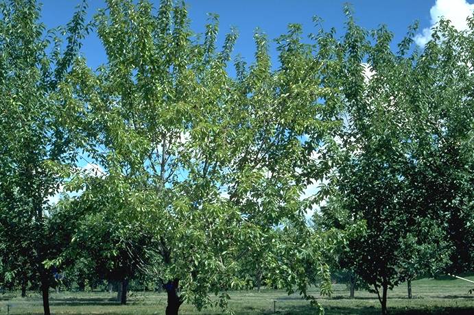
- 226 Sweet_cherry,_X-disease_(MLO),_tree_decline_(Mazzard_rootstock)
- 227 Sweet_cherry_&_peach,_cherry_rasp-leaf,_symptoms_on_leaves
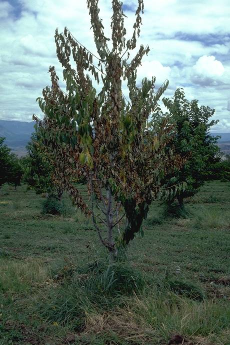
- 228 Tart_cherry,_X-disease,_tree_collapse_(Mahaleb_rootstock)
- 229 Tomato_bushy_stunt_virus
- 230 Tomato_ringspot_virus
- 231 Tomato_ringspot_virus
- 232 Tomato_ringspot_virus
- 233 Verticillium_Wilt_1
- 234 Verticillium_Wilt_2
- 235 X-Disease_1
- 236 X-Disease_2
- 237 X-Disease_3
- 238 X-Disease_4
- 239 X-Disease_5
- 240 X-Disease_6
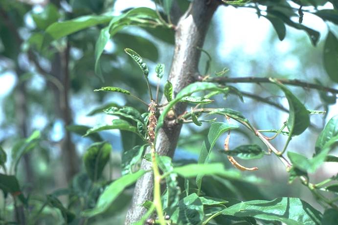
- 241 X-Disease_7
Alternaria_Fruit_Rot_1
Alternaria Fruit Rot 1 ==
Alternaria rot on dark sweet cherry fruit. Note flattened and wrinkled nature of the lesions.
(Alan L. Jones, Michigan State University and Turner B. Sutton, North Carolina State University)

Alternaria_Fruit_Rot_2
Alternaria Fruit Rot 2 ==
Decay of Gold sweet cherry fruit due to Alternaria rot.
(Alan L. Jones, Michigan State University and Turner B. Sutton, North Carolina State University)
American_Brown_Rot_1
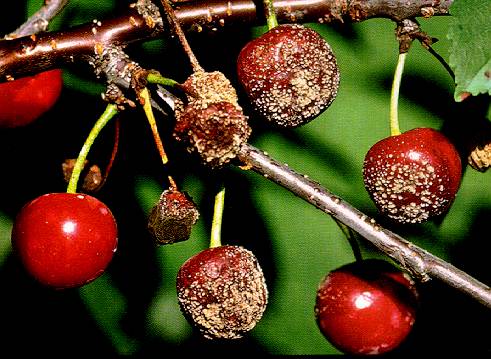
American Brown Rot 1 ==
Decay of sour cherries by Monilinia fructicola and sporulation of the pathogen.
(Alan L. Jones, Michigan State University and Turner B. Sutton, North Carolina State University)
American_Brown_Rot_2
American Brown Rot 2 ==
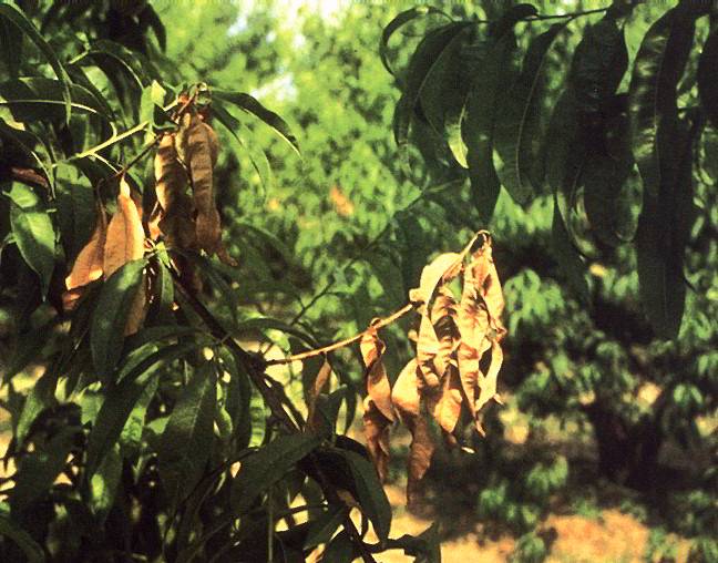
Decay of peach and dieback of the shoot growth from brown rot.
(Alan L. Jones, Michigan State University and Turner B. Sutton, North Carolina State University)
American_Brown_Rot_3
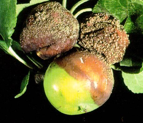
American Brown Rot 3 ==
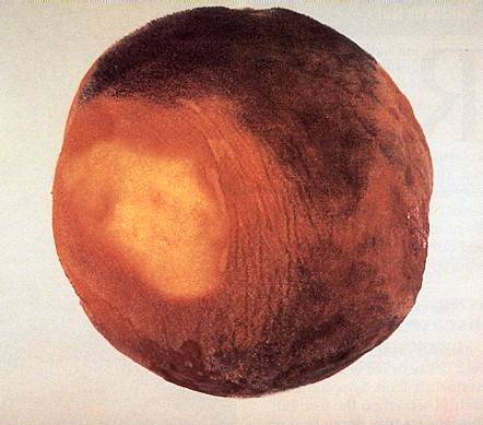
Brown rot on apple.
(Alan L. Jones, Michigan State University and Turner B. Sutton, North Carolina State University)
American_Brown_Rot_4
American Brown Rot 4 ==
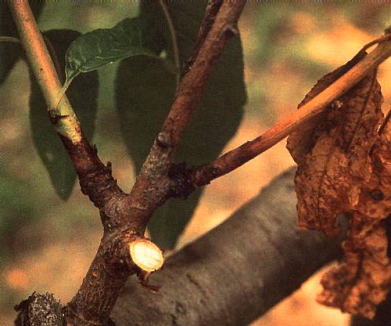
Sporulation of the brown rot fungus on peach following infection of the shoot tip.
(Alan L. Jones, Michigan State University and Turner B. Sutton, North Carolina State University)
American_Brown_Rot_5
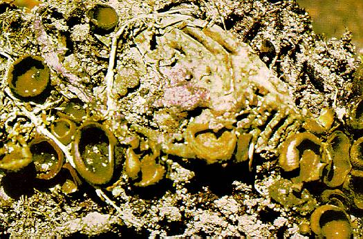
American Brown Rot 5 ==
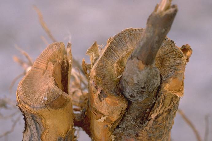
Apothecia or cup-shaped fruiting bodies of brown rot arising from a peach mummy.
(Alan L. Jones, Michigan State University and Turner B. Sutton, North Carolina State University)
Anthracnose_1a Anthracnose 1a
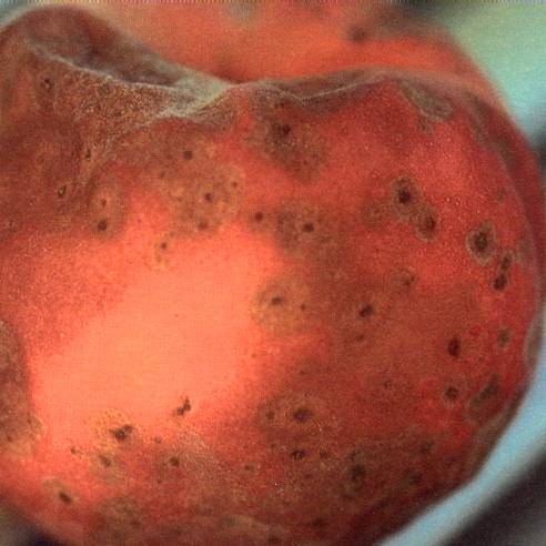
Anthracnose lesions on peach. Initial infection, later with concentric rings of acervuli and salmon spores.
(Alan L. Jones, Michigan State University and Turner B. Sutton, North Carolina State University)
Anthracnose_1b
Anthracnose 1b ==
Anthracnose lesions on peach -- with concentric rings of acervuli and salmon spores.
(Alan L. Jones, Michigan State University and Turner B. Sutton, North Carolina State University)
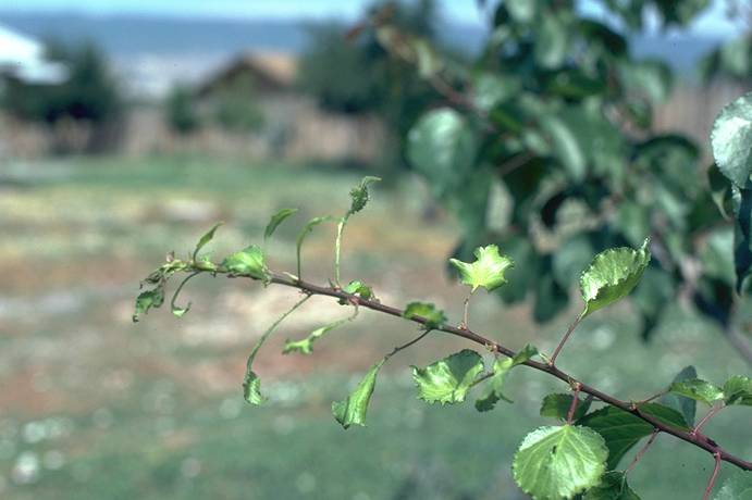
Apricot,_2,4-D_herbicide_injury
Apricot, 2,4-D herbicide injury (A. R. Renquist; Colorado State University)
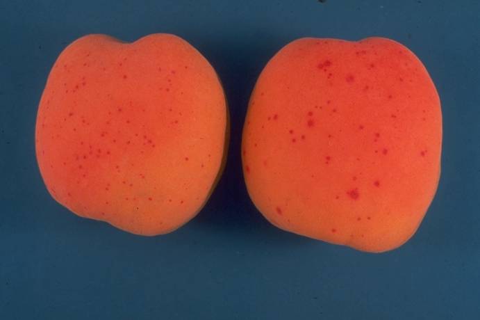
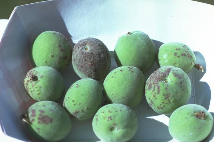
Apricot,_Alternaria_spot,_fruit_symptoms
Apricot, Alternaria spot, fruit symptoms (H. J. Larsen; Colorado State University)
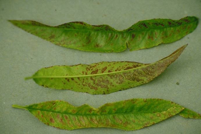
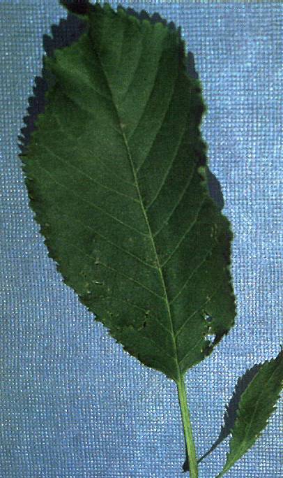
Apricot,_asteroid_spot,_leaf_symptoms
Apricot, asteroid spot, leaf symptoms (N. S. Luepschen; Colorado State University)
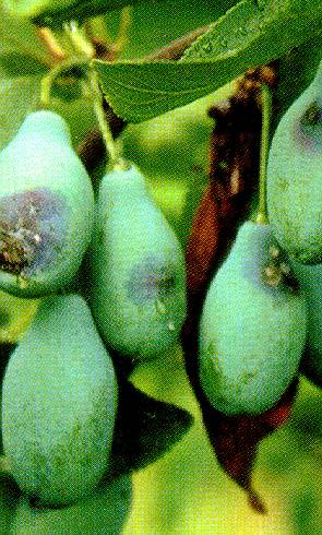
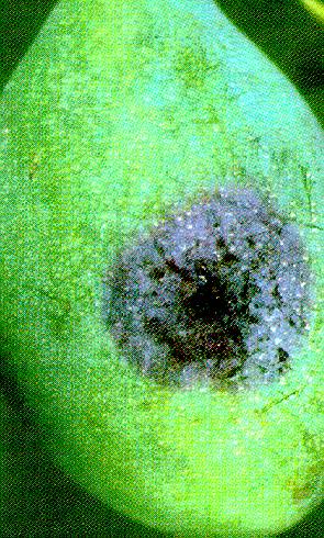
Apricot,_Coryneum_fruit_infection
Apricot, Coryneum fruit infection (N. S. Luepschen; Colorado State University)
Apricot,_hail_damage,_fruit
Apricot, hail damage, fruit (C. R. Ure; Colorado State University)
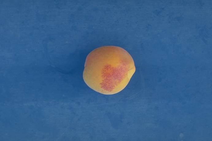
Apricot,_powdery_mildew,_russet
Apricot, powdery mildew, russet (H. J. Larsen; Colorado State University)
Apricot,_Roundup_herbicide_damage
Apricot, Roundup herbicide damage (A. R. Renquist; Colorado State University)
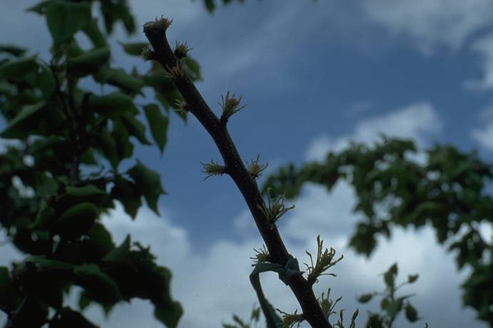
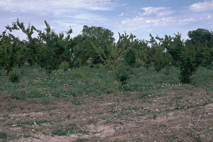
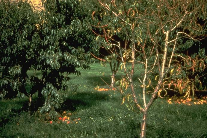
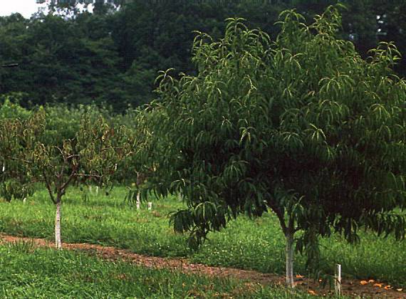
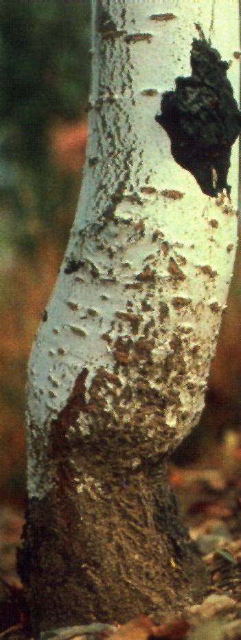
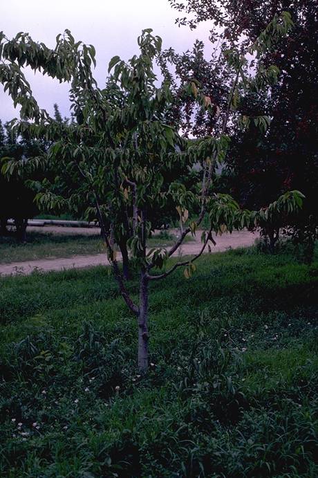
Apricot_decline,_on_plum_rootstock_(incompatibility)
Apricot decline, on plum rootstock (incompatibility)
(H. J. Larsen; Colorado State University)
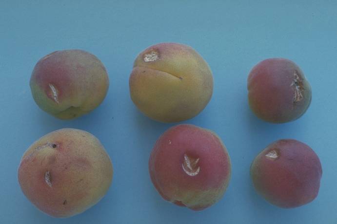
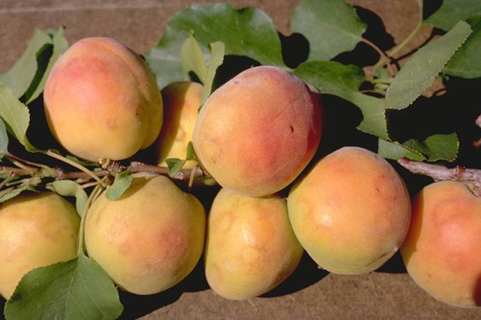
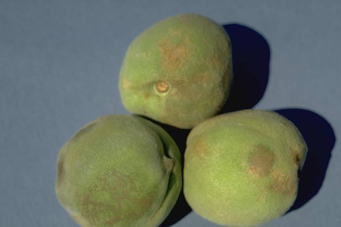
Apricot_ring_pox_virus_on_fruit
Apricot ring pox virus on fruit (N. S. Luepschen; Colorado State University)
Apricot_ring_pox_virus_on_fruit_
Apricot ring pox virus on fruit (N. S. Luepschen; Colorado State University)
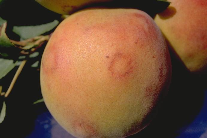
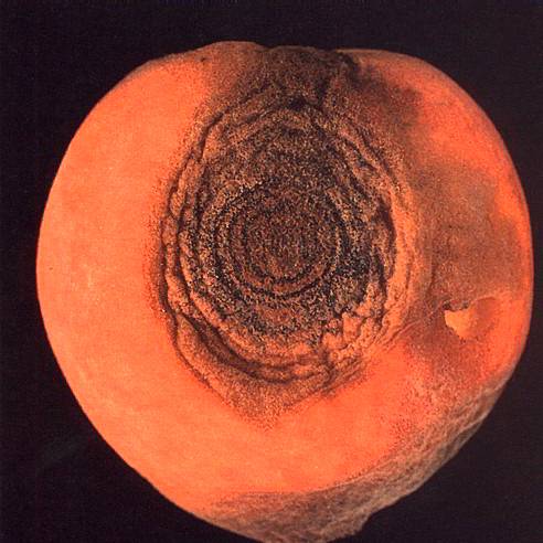
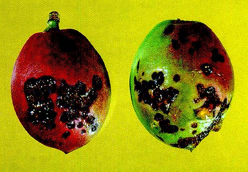
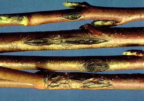
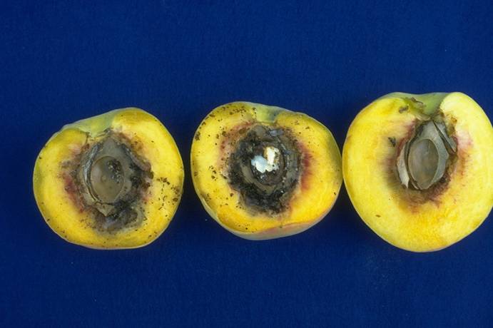
Apricot_ring_pox_virus_on_fruit_(lesions)
Apricot ring pox virus on fruit (lesions)
(H. J. Larsen; Colorado State University)
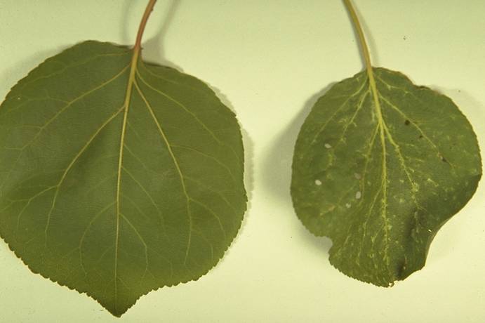
Apricot_ring_pox_virus_on_leaves_(red_blotched_veins)
Apricot ring pox virus on leaves (red blotched veins)
(H. J. Larsen; Colorado State University)

Armillaria_and_Clitocybe_Root_Rots_1
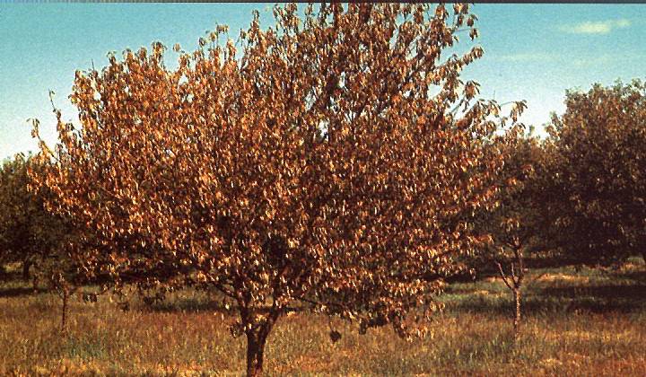
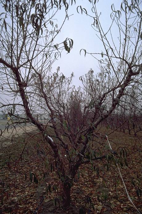
Armillaria and Clitocybe Root Rots 1 ==
Sudden collapse of a Montmorency sour cherry tree in midsummer caused by Armillaria root rot. Leaves remain attached into winter.
(Alan L. Jones, Michigan State University and Turner B. Sutton, North Carolina State University)
Armillaria_and_Clitocybe_Root_Rots_2a
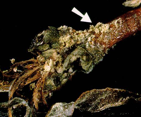
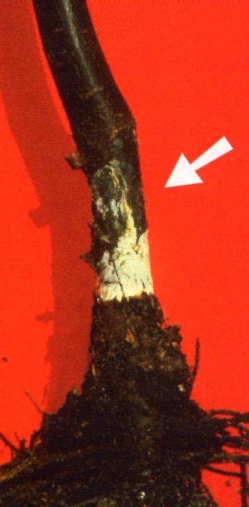
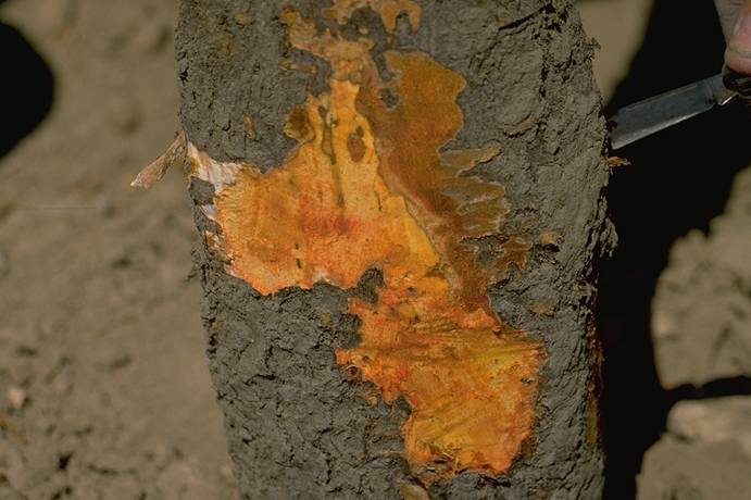
Armillaria and Clitocybe Root Rots 2a ==
Armillaria root rot. White fungal mat beneath the bark and necrotic tissue with white fungal growth beneath the bark.
(Alan L. Jones, Michigan State University and Turner B. Sutton, North Carolina State University)
Armillaria_and_Clitocybe_Root_Rots_2b
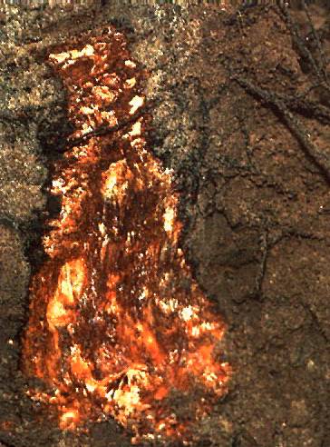
Armillaria and Clitocybe Root Rots 2b ==
Armillaria root rot -- rhizomorphs in the surrounding soil right.
(Alan L. Jones, Michigan State University and Turner B. Sutton, North Carolina State University)
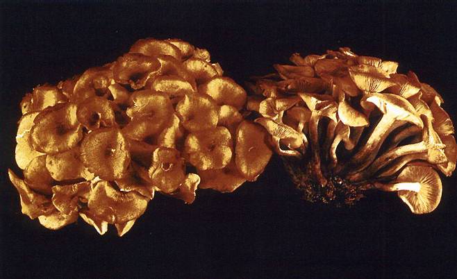
Armillaria_and_Clitocybe_Root_Rots_3
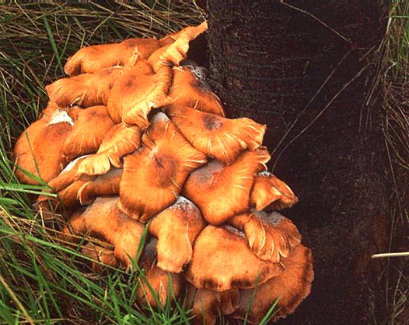
Armillaria and Clitocybe Root Rots 3 ==
Mushrooms of Armillaria ostoyae around the base of a cherry trunk.
(Alan L. Jones, Michigan State University and Turner B. Sutton, North Carolina State University)
Armillaria_and_Clitocybe_Root_Rots_4
Armillaria and Clitocybe Root Rots 4 ==
The mushrooms of Armillaria tabescens lack an annulus around the stripe.
(Alan L. Jones, Michigan State University and Turner B. Sutton, North Carolina State University)
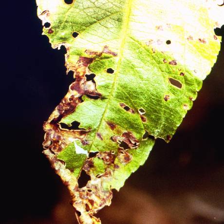
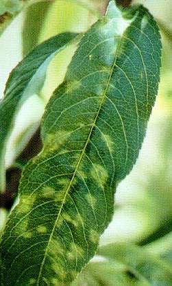
Bacteral_Spot_of_Leaf
Bacteral Spot of Leaf
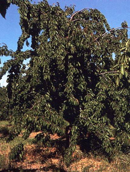
Bacterial_Canker_1a
Bacterial Canker 1a ==
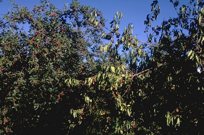
Healthy Hardy Giant sweet cherry tree on left (see infected tree).
(Alan L. Jones, Michigan State University and Turner B. Sutton, North Carolina State University)
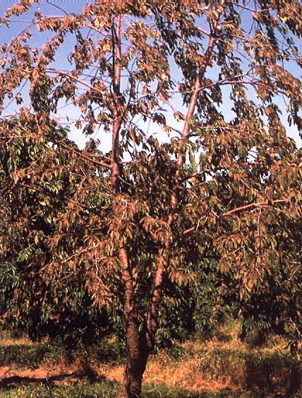
Bacterial_Canker_1b
Bacterial Canker 1b ==
Hardy Giant sweet cherry tree with systemic bacterial canker infection on right. Note upright growth and discoloration of infected tree.
(Alan L. Jones, Michigan State University and Turner B. Sutton, North Carolina State University)
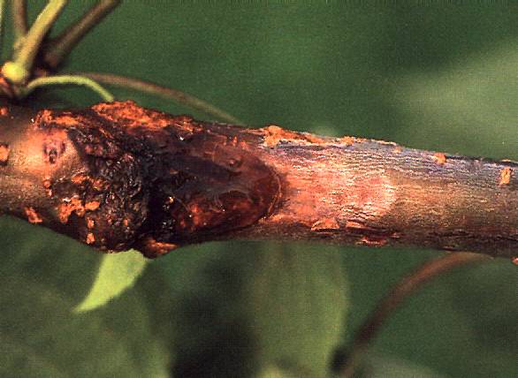
Bacterial_Canker_2
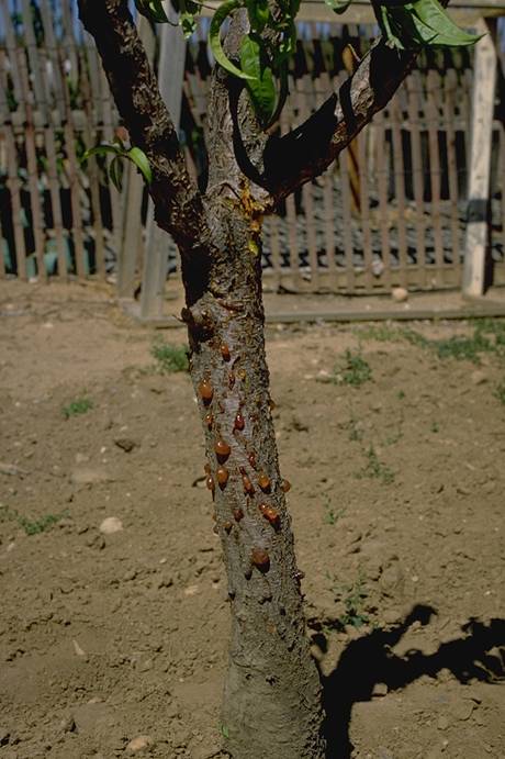
Bacterial Canker 2 ==
Gum exudation and upward extension of a canker on sweet cherry caused by Pseudomonas syringae pv. morsprunorum.
(Alan L. Jones, Michigan State University and Turner B. Sutton, North Carolina State University)
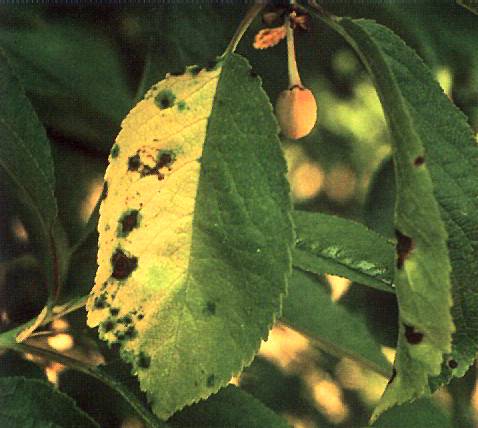
Bacterial_Canker_3
Bacterial Canker 3 ==
Sour cherry with necrotic spots and yellowing of leaves due to bacterial canker.
(Alan L. Jones, Michigan State University and Turner B. Sutton, North Carolina State University)
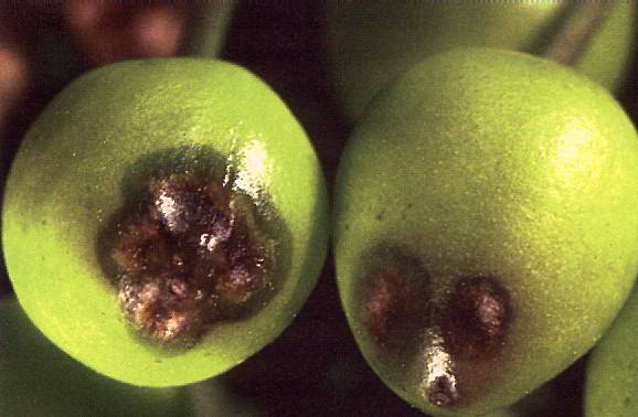
Bacterial_Canker_4
Bacterial Canker 4 ==
Water soaking around necrotic lesions on immature cherry fruit caused by bacterial canker.
(Alan L. Jones, Michigan State University and Turner B. Sutton, North Carolina State University)
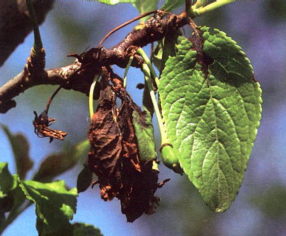
Bacterial_Canker_5
Bacterial Canker 5 ==
Spur dieback and necrosis of the midvein of Stanley plum caused by bacterial canker.
(Alan L. Jones, Michigan State University and Turner B. Sutton, North Carolina State University)
Bacterial_Canker_6
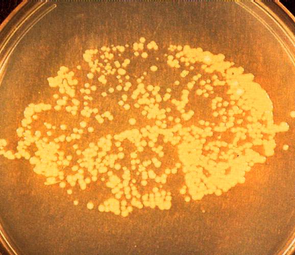
Bacterial Canker 6 ==
Leaf print made by colonies of the bacterial canker organism on a culture medium demonstrates the presence of bacteria on the surface of an apparently healthy leaf.
(Alan L. Jones, Michigan State University and Turner B. Sutton, North Carolina State University)
Bacterial_Spot_1
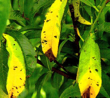
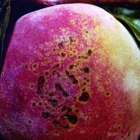
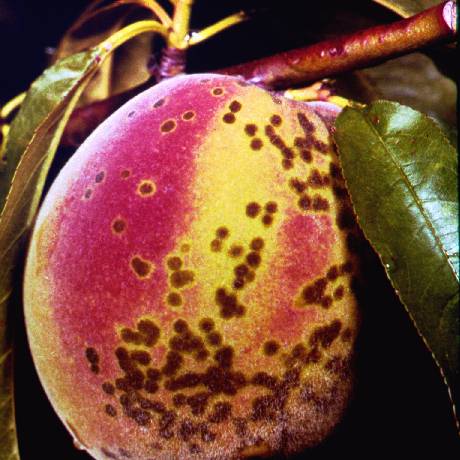
Bacterial Spot 1 ==
Yellowing and tip burning of peach leaves with bacterial spot, caused by Xanthomonas campestris pv. pruni,
(Alan L. Jones, Michigan State University and Turner B. Sutton, North Carolina State University)
Bacterial_Spot_2
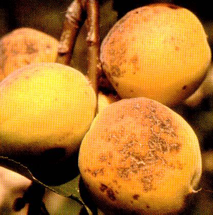
Bacterial Spot 2 ==
Pitting of the flesh of peach due to bacterial spot.
(Alan L. Jones, Michigan State University and Turner B. Sutton, North Carolina State University)
Bacterial_Spot_3
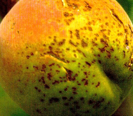
Bacterial Spot 3 ==
Bacterial spot on apricot.
(Alan L. Jones, Michigan State University and Turner B. Sutton, North Carolina State University)
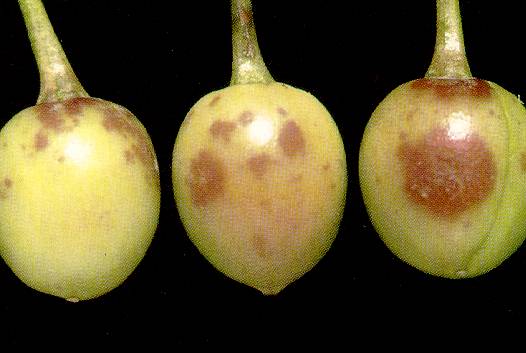
Bacterial_Spot_4a
Bacterial Spot 4a ==
Stanley plum with bacterial spot. Note purple lesions.
(Alan L. Jones, Michigan State University and Turner B. Sutton, North Carolina State University)
Bacterial_Spot_4b
Bacterial Spot 4b ==
Stanley plum with bacterial spot. Note purple lesions.
(Alan L. Jones, Michigan State University and Turner B. Sutton, North Carolina State University)
Bacterial_Spot_5
Bacterial Spot 5 ==
Pitting and gumming on nectarines with bacterial spot.
(Alan L. Jones, Michigan State University and Turner B. Sutton, North Carolina State University)
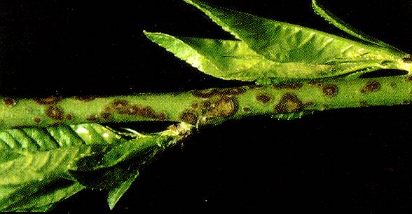
Bacterial_Spot_6
Bacterial Spot 6 ==
Bacterial spot cankers on peach at bud break in spring.
(Alan L. Jones, Michigan State University and Turner B. Sutton, North Carolina State University)
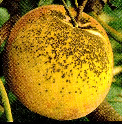
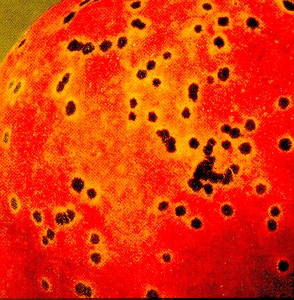
Bacterial_Spot_on_Fruit
Bacterial Spot on Fruit
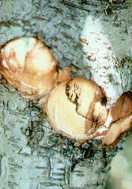
Bing_cherry,_X-disease,_graft_union_symptoms_(Mahaleb_rootstock)
Bing cherry, X-disease, graft union symptoms (Mahaleb rootstock)
(H. J. Larsen; Colorado State University)
Black_Knot_of_Plum_-_Plum
Black Knot of Plum - Plum
Black_Knot_of_Plum_1a
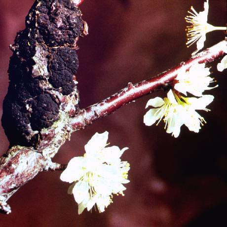
Black Knot of Plum 1a ==
Young knots on plum shoots caused by black knot.
(Alan L. Jones, Michigan State University and Turner B. Sutton, North Carolina State University)
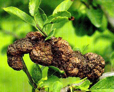
Black_Knot_of_Plum_1b
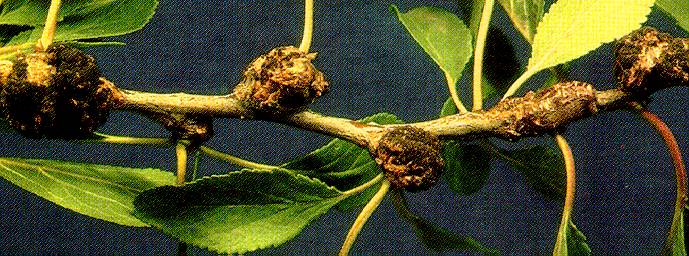
Black Knot of Plum 1b ==
Old knots on plum shoots caused by black knot.
(Alan L. Jones, Michigan State University and Turner B. Sutton, North Carolina State University)
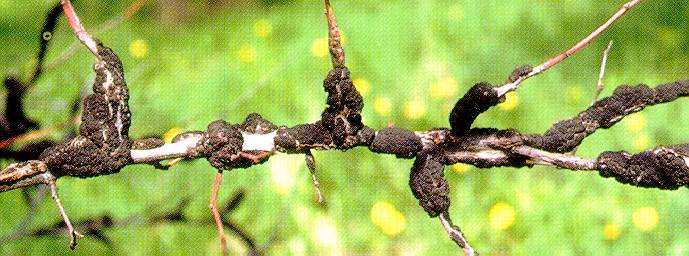
Black_Knot_of_Plum_2
Black Knot of Plum 2 ==
Black knot lesion on plum after about 3 years.
(Alan L. Jones, Michigan State University and Turner B. Sutton, North Carolina State University)
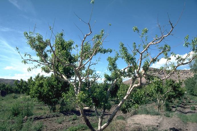
Blast_Disease_on_Trunk
Blast Disease on Trunk
Blossom_Blight
Blossom Blight
Brown_Rot_on_Fruit
Brown Rot on Fruit
Cherry,_Coryneum_leaf_infection
Cherry, Coryneum leaf infection (N. S. Luepschen; Colorado State University)
Cherry,_2,4-D_herbicide_injury
Cherry, 2,4-D herbicide injury (N. S. Luepschen; Colorado State University)
Cherry_Leaf_Spot_1
Cherry Leaf Spot 1 ==
Cherry leaf spot infection on sweet cherry leaves.
(Alan L. Jones, Michigan State University and Turner B. Sutton, North Carolina State University)
Cherry_Leaf_Spot_2a
Cherry Leaf Spot 2a ==
Sporulation of the pathogen in acervuli on the lower surface caused by cherry leaf spot.
(Alan L. Jones, Michigan State University and Turner B. Sutton, North Carolina State University)

Cherry_Leaf_Spot_2b
Cherry Leaf Spot 2b ==
Sporulation of the pathogen in acervuli, circular spots plus yellowing of sour cherry leaves (right) caused by cherry leaf spot.
(Alan L. Jones, Michigan State University and Turner B. Sutton, North Carolina State University)
Cherry_Leaf_Spot_3
Cherry Leaf Spot 3 ==
Defoliation by cherry leaf spot of unsprayed trees (right) compared with no defoliation on fungicide-sprayed tree(left).
(Alan L. Jones, Michigan State University and Turner B. Sutton, North Carolina State University)
Cherry_Leaf_Spot_4
Cherry Leaf Spot 4 ==
Stems of sour cherry fruit with sporulating cherry leaf spot lesions. Note single lesions on the two right fruit.
(Alan L. Jones, Michigan State University and Turner B. Sutton, North Carolina State University)
Cherry_Leaf_Spot_5
Cherry Leaf Spot 5 ==
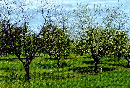
Death of cherry tree on left in winter following severe defoliation from leaf spot the previous summer. Leaf spot was controlled on the tree that is in bloom.
(Alan L. Jones, Michigan State University and Turner B. Sutton, North Carolina State University)
Cherry_Leaf_Spot_6
Cherry Leaf Spot 6 ==
Apothecia of the leaf spot fungus on overwintered sweet cherry leaves.
(Alan L. Jones, Michigan State University and Turner B. Sutton, North Carolina State University)
Cherry_Leaf_Spot_7a
Cherry Leaf Spot 7a ==
Asci with ascospores of the cherry leaf spot fungus, Blumeriella jaapii.
(Alan L. Jones, Michigan State University and Turner B. Sutton, North Carolina State University)

Cherry_Leaf_Spot_7b
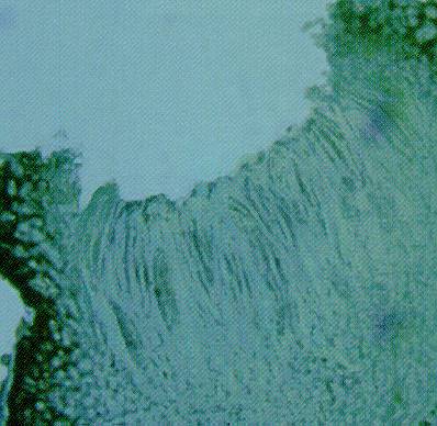
Cherry Leaf Spot 7b ==
Asci with cross-section of an apothecium of the cherry leaf spot fungus, Blumeriella jaapii.
(Alan L. Jones, Michigan State University and Turner B. Sutton, North Carolina State University)
Cherry_Leaf_Spot_8
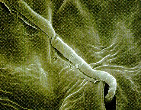
Cherry Leaf Spot 8 ==
Germinated conidium of the leaf spot fungus infecting through a stomate on the underside of a sour cherry leaf.
(Alan L. Jones, Michigan State University and Turner B. Sutton, North Carolina State University)
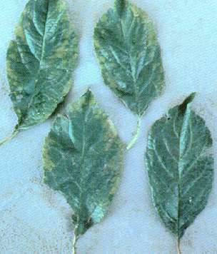
Cherry_mottle_leaf_virus
Cherry mottle leaf virus (H. J. Larsen; Colorado State University)
Cherry_mottle_leaf_virus_on_sweet_cherry
Cherry mottle leaf virus on sweet cherry (H. J. Larsen; Colorado State University)
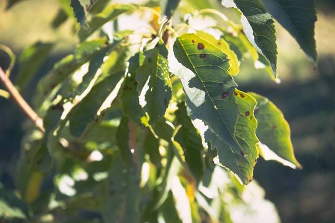
Cherry_rusty_mottle_disease
Cherry rusty mottle disease leaf symptoms (pitfruit, stone fruit, disease) (J.A. Foster, USDA)
Cherry_stem_pitting_(virus),_pitting_symptoms_at_graft_union
Cherry stem pitting (virus), pitting symptoms at graft union (S. Savage; Colorado State University)
Cherry_stem_pitting(virus),_whole_tree_collapse
Cherry stem pitting (virus), whole tree collapse (S. Savage; Colorado State University)
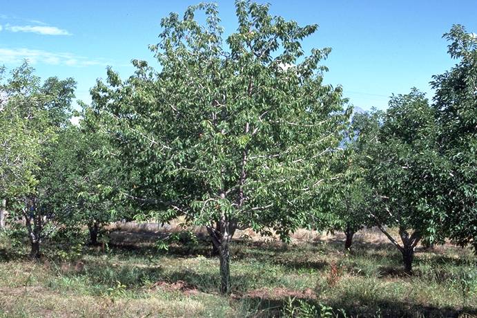
Cherry_tree,_weak
Cherry tree, weak (S. Savage; Colorado State University)
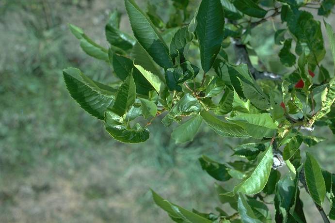
Cherry_twisted_leaf_virus,_leaf_symptoms
Cherry twisted leaf virus, leaf symptoms (H. J. Larsen; Colorado State University)
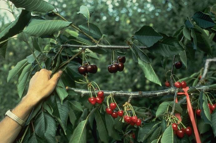
Cherry_X-disease_(little_cherry),_symptomatic_vs_normal
Cherry X-disease (little cherry), symptomatic vs normal (C. L. Parish; Colorado State University)
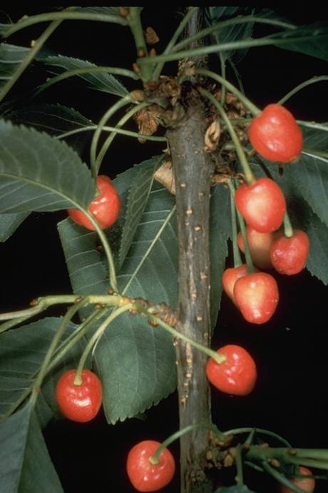
Cherry_X-disease_(little_cherry)_fruit_symptoms_on_Lambert
Cherry X-disease (little cherry) fruit symptoms on Lambert (C. L. Parish; Colorado State University)
Cherry_X-disease_(little_cherry)_leaf_symptoms
Cherry X-disease (little cherry) leaf symptoms (C. L. Parish; Colorado State University)
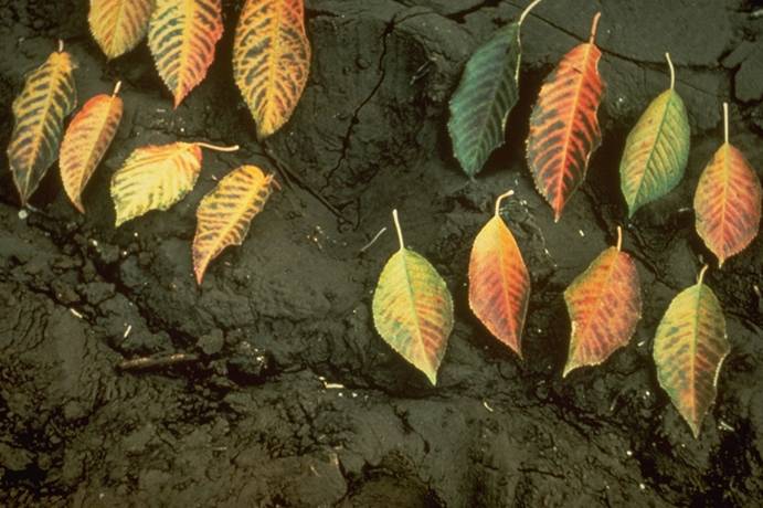
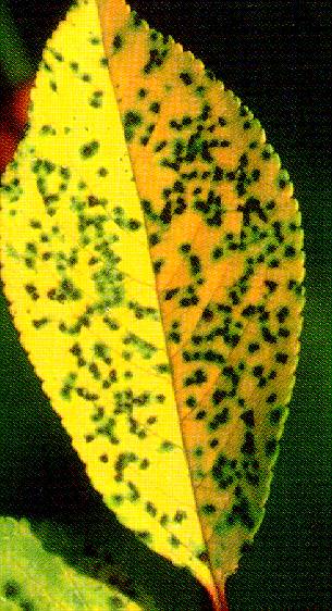
Chokecherry,_1st_year_symptoms_on_leaves
Chokecherry, 1st year symptoms on leaves (C. L. Parish; Colorado State University)
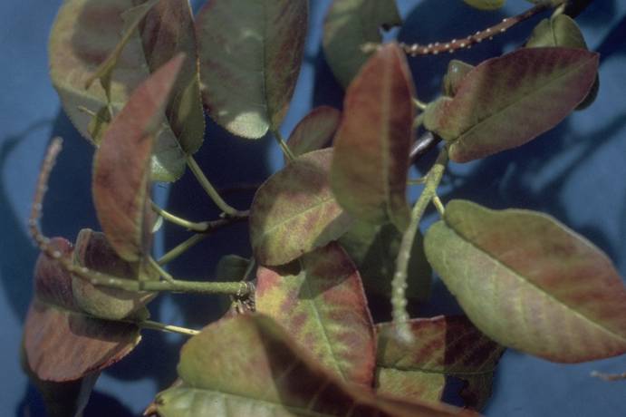
Chokecherry,_X-disease,_early_leaf_coloration_symptoms
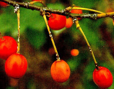
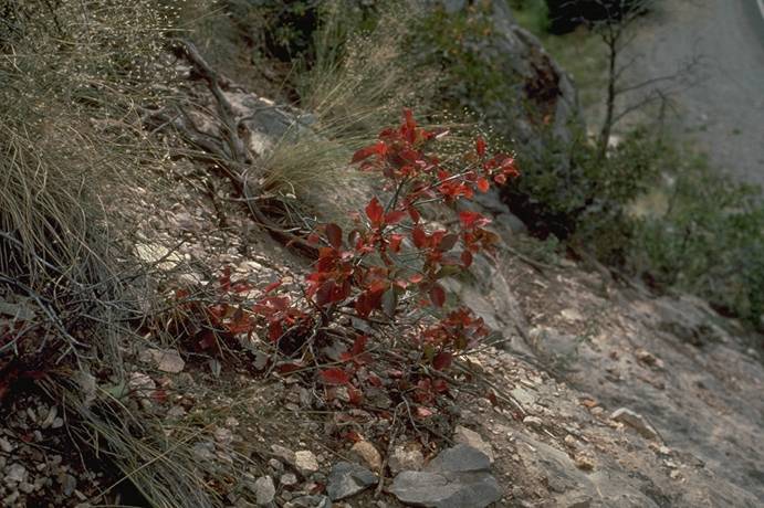
Chokecherry, X-disease, early leaf coloration symptoms (H. J. Larsen; Colorado State University)
Chokecherry,_X-disease,_early_leaf_coloration_symptoms
Chokecherry, X-disease, early leaf coloration symptoms (H. J. Larsen; Colorado State University)
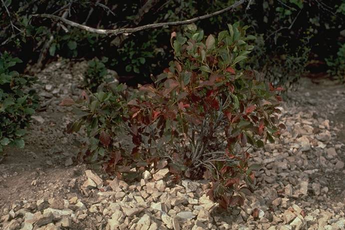
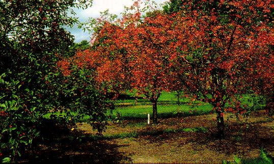
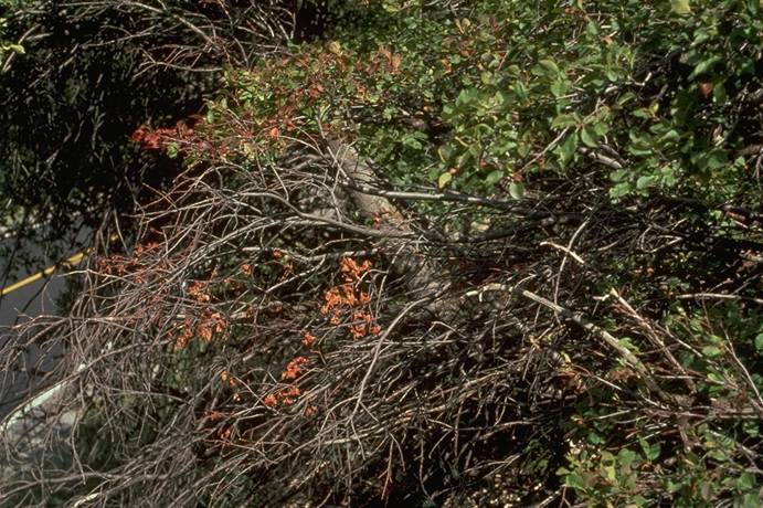
Chokecherry,_X-disease,_symptoms
Chokecherry, X-disease, symptoms (H. J. Larsen; Colorado State University)
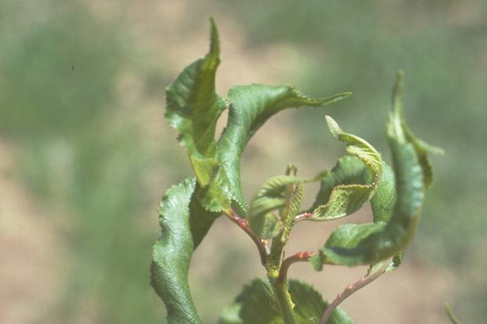
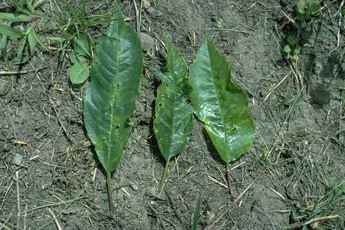
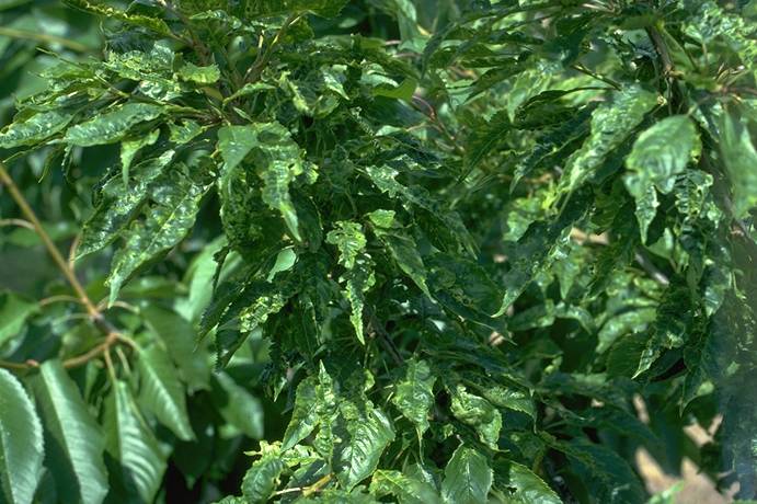
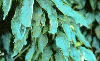
Crinkle_leaf
Bing sweet cherry, crinkle leaf affected leaves: deformed, irregularly indented, scattered areas along leaf margins, chlorotic, light green (pitfruit, stone fruit, disease) (J.K. Uyemoto, USDA)
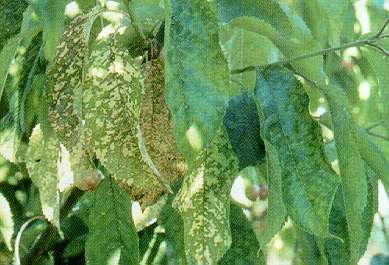
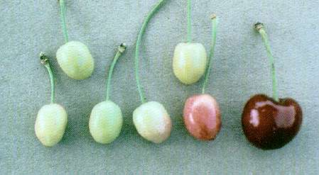
Crinkle_leaf
Small fruit symptoms, cherry crinkle leaf: fruit pedicels vary, length; healthy fruit, far right (pitfruit, stone fruit, disease) (J.K. Uyemoto, USDA)
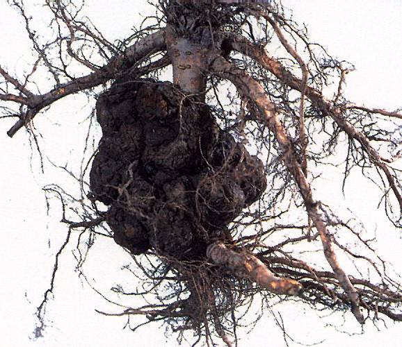
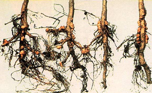
Crown_Gall_1
Crown Gall 1 ==
Crown gall of Mazzard F12/1 rootstock.
(Alan L. Jones, Michigan State University and Turner B. Sutton, North Carolina State University)
Crown_Gall_2a
Crown Gall 2a ==
Biological control of crown gall on Prunus seedlings with Agrobacterium radiobacter strain 84.
(Alan L. Jones, Michigan State University and Turner B. Sutton, North Carolina State University)
Crown_Gall_2b Crown Gall 2b
Biological control of crown gall on Prunus seedlings with Agrobacterium radiobacter strain 84. Treated seedlings were dipped in a solution of strain 84, then after 24 hours both sets of seedlings were inoculated with Agrobacterium tumefaciens, the crown gall pathogen.
(Alan L. Jones, Michigan State University and Turner B. Sutton, North Carolina State University)

Cycle_brown_rot
Cycle brown rot ==
Disease cycle of brown rot.
(Alan L. Jones, Michigan State University and Turner B. Sutton, North Carolina State University)

Cycle_cherry_leaf_spot
Cycle cherry leaf spot ==
Disease cycle of cherry leaf spot.
(Alan L. Jones, Michigan State University and Turner B. Sutton, North Carolina State University)

Cycle_peach_bacterial_spot
Cycle peach bacterial spot ==
Disease cycle of peach bacterial spot.
(Alan L. Jones, Michigan State University and Turner B. Sutton, North Carolina State University)

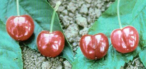
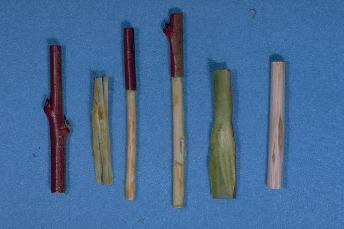
Deep_suture_symptoms
Bing cherry fruit, deep suture symptoms shown on right: indentation along, crease (suture line); two healthy fruit, left (pitfruit, stone fruit, disease) (J.K. Uyemoto, USDA)
Early_Red_Haven_peach,_peach_mosaic_rosetting_on_right
Early Red Haven peach, peach mosaic rosetting on right (H. J. Larsen; Colorado State University)
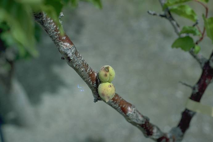
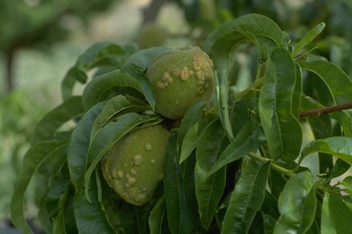
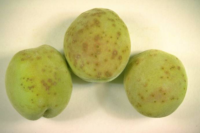
Elberta_peach,_peach_wart,_fruit_symptoms
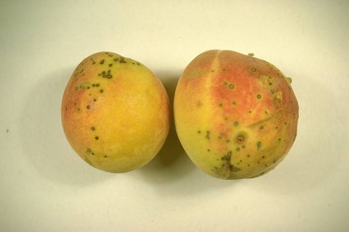
Elberta peach, peach wart, fruit symptoms (H. J. Larsen; Colorado State University)
Elberta_peach,_peach_wart,_fruit_symptoms
Elberta peach, peach wart, fruit symptoms (H. J. Larsen; Colorado State University)
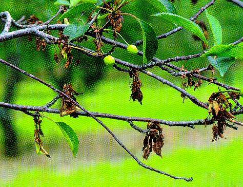
European_Brown_Rot_1
European Brown Rot 1 ==
Spur dieback of Montmorency sour cherry caused by the European brown rot fungus. Monilinia laxa.
(Alan L. Jones, Michigan State University and Turner B. Sutton, North Carolina State University)
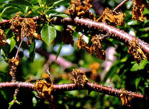
European_Brown_Rot_2
European Brown Rot 2 ==
Spur dieback of Meteor sour cherry caused by European brown rot.
(Alan L. Jones, Michigan State University and Turner B. Sutton, North Carolina State University)
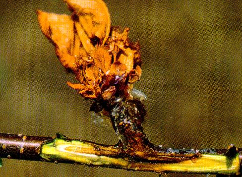
European_Brown_Rot_3
European Brown Rot 3 ==
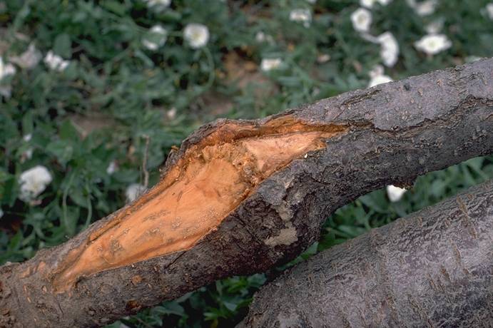
Canker and gumming around a fruiting spur killed by European brown rot. Bark was removed to reveal canker.
(Alan L. Jones, Michigan State University and Turner B. Sutton, North Carolina State University)
Fusicoccum_Canker_1
Fusicoccum Canker 1 ==
Death of peach shoots caused by Fusicoccum canker.
(Alan L. Jones, Michigan State University and Turner B. Sutton, North Carolina State University)
Fusicoccum_Canker_2
Fusicoccum Canker 2 ==
Close-up of constriction cankers at the base of shoots blighted by Phomopsis amygdali. The cankers result from toxins produced by the pathogen.
(Alan L. Jones, Michigan State University and Turner B. Sutton, North Carolina State University)
Gibertella_Rot
Gibertella Rot ==
Gilbertella rot of peach.
(Alan L. Jones, Michigan State University and Turner B. Sutton, North Carolina State University)
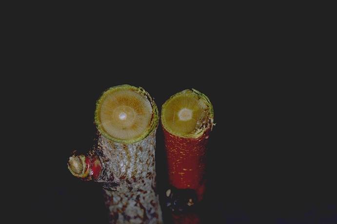
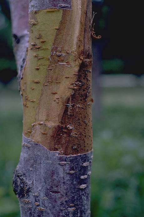
Graft_union_incompatibility_(TmRS)
Graft union incompatibility (TmRS)
(H. J. Larsen; Colorado State University)
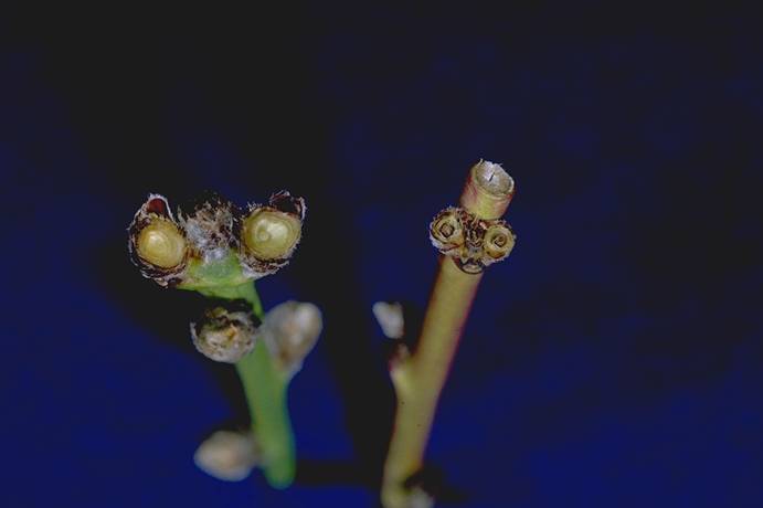
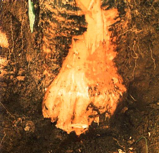
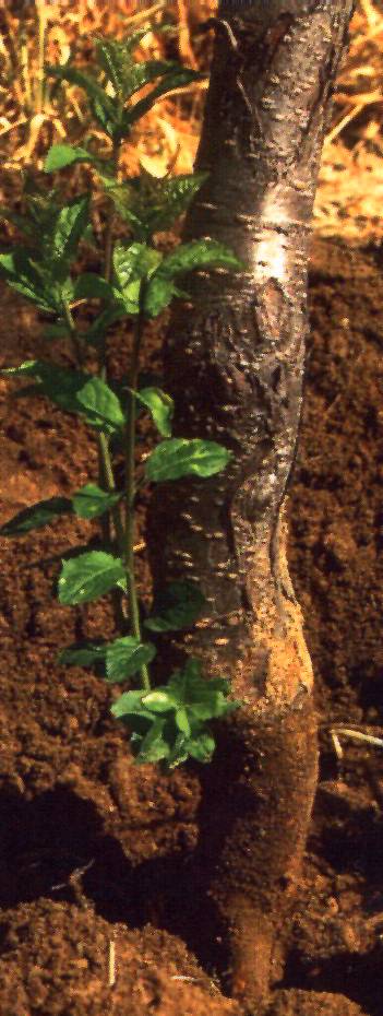
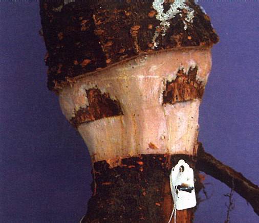
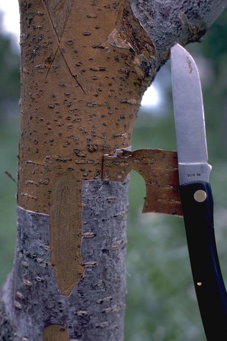
Graft_union_of_apricot_decline_on_plum_rootstock
Graft union of apricot decline on plum rootstock (H. J. Larsen; Colorado State University)
Green_Ring_Mottle_1
Green Ring Mottle 1 ==
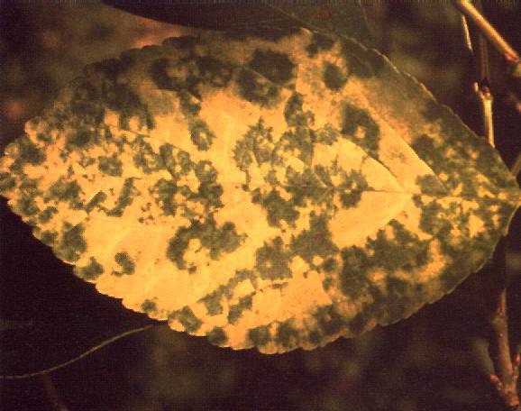
Dark green arcs and rings on yellow background in Montmorency cherry leaves are typical of green ring mottle.
(Alan L. Jones, Michigan State University and Turner B. Sutton, North Carolina State University)
Green_Ring_Mottle_2
Green Ring Mottle 2 ==
Constricting chlorosis symptoms on Montmorency cherry leaves caused by green ring mottle.
(Alan L. Jones, Michigan State University and Turner B. Sutton, North Carolina State University)
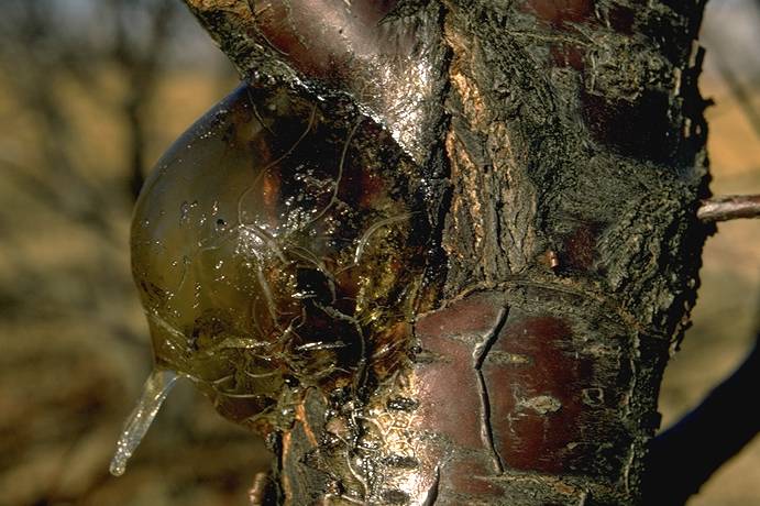
Gummosis
Gummosis ==
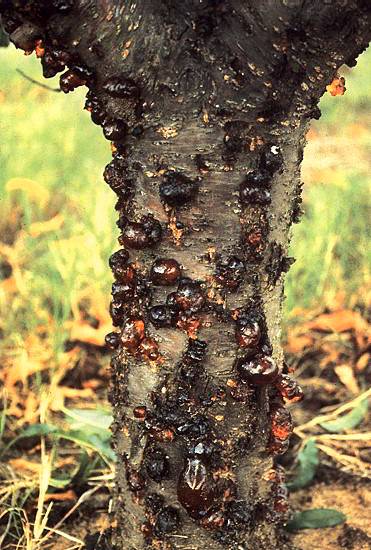
Extensive exudation of gum on the trunk of peach (gummosis) caused by Botryosphaeria spp.
(Alan L. Jones, Michigan State University and Turner B. Sutton, North Carolina State University)
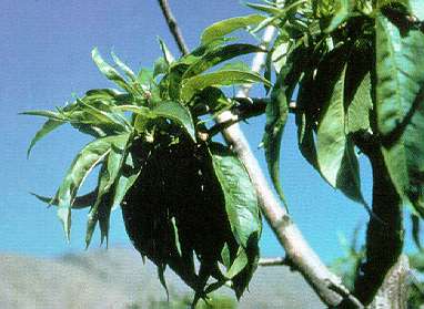
Lambert_sweet_cherry,_little_cherry_virus,_fruit_symptoms
Lambert sweet cherry, little cherry virus, fruit symptoms (H. J. Larsen; Colorado State University)
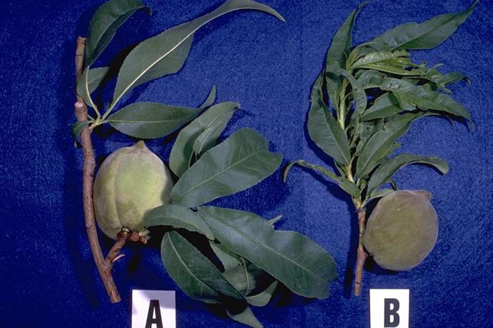
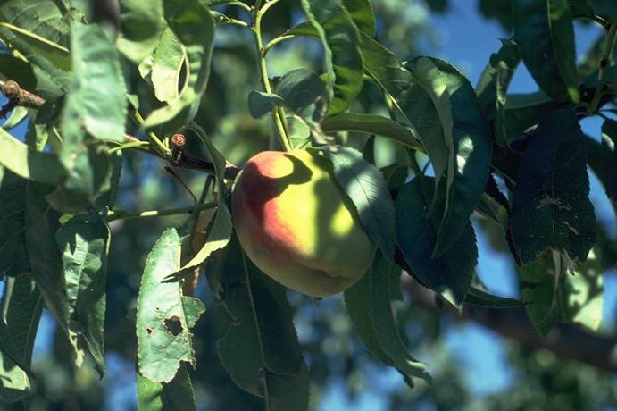
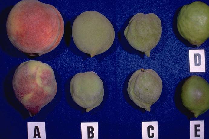
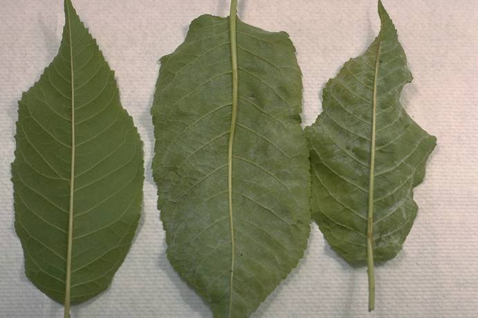
Loring_peach,_A._healthy,_B._symptomatic
Loring peach, A. healthy, B. symptomatic (H. J. Larsen; Colorado State University)
Montmorency_sour_cherry_+_sap_exudation,_ethrel_treated_@_2000_ppm
Montmorency sour cherry + sap exudation, ethrel treated @ 2000 ppm (N. S. Luepschen; Colorado State University)
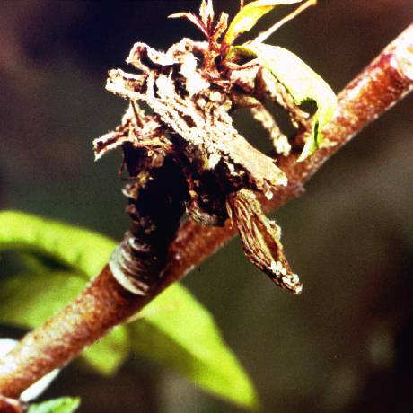
Morello_sour_cherry,_crown_borer_injury
Morello sour cherry, crown borer injury (N. S. Luepschen; Colorado State University)
Morello_sour_cherry,_crown_borer_injury_on_right
Morello sour cherry, crown borer injury on right (N. S. Luepschen; Colorado State University)
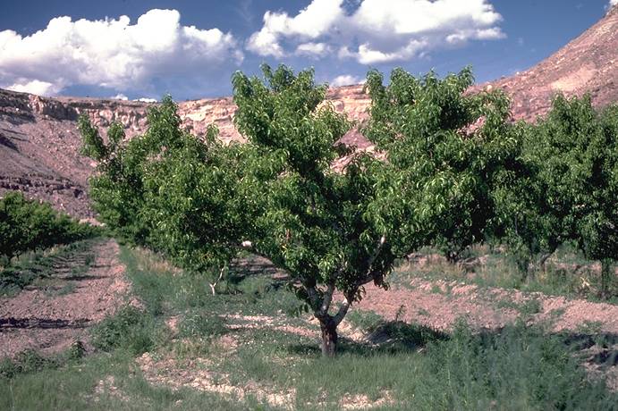
Orchard_Disease_Problem
Orchard Disease Problem
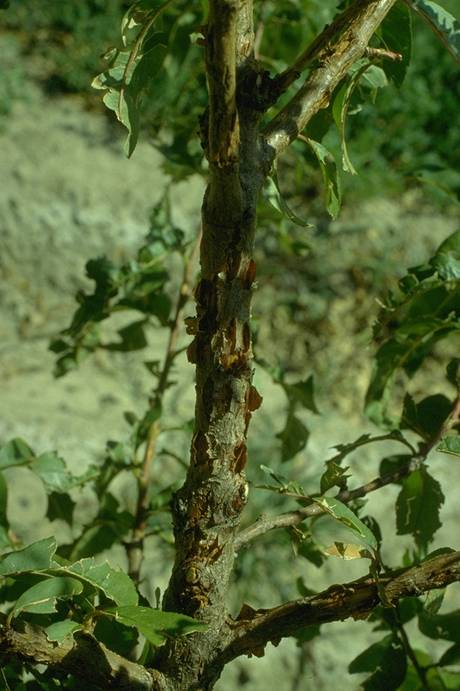
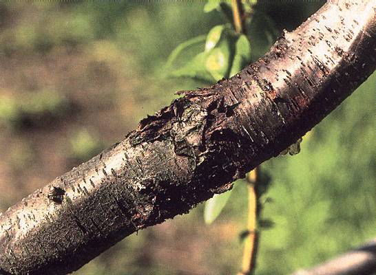
Peach,_Botryosphaeria_canker,_bark_tissue_symptoms
Peach, Botryosphaeria canker, bark tissue symptoms (H. J. Larsen; Colorado State University)
Peach,_Botryosphaeria_canker,_killed_tree_with_severe_gumming
Peach, Botryosphaeria canker, killed tree with severe gumming (H. J. Larsen; Colorado State University)
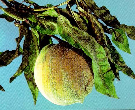
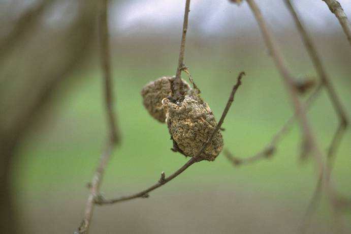
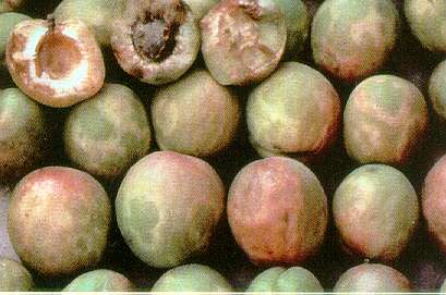
Peach,_brown_rot_mummy
Peach, brown rot mummy (H. J. Larsen; Colorado State University)
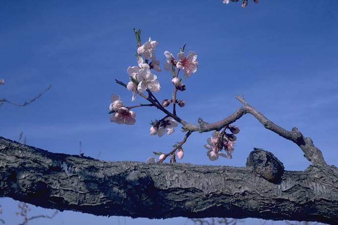
Peach,_burr_knot_(aerial_crown_gall)
Peach, burr knot (aerial crown gall)
(N. S. Luepschen; Colorado State University)
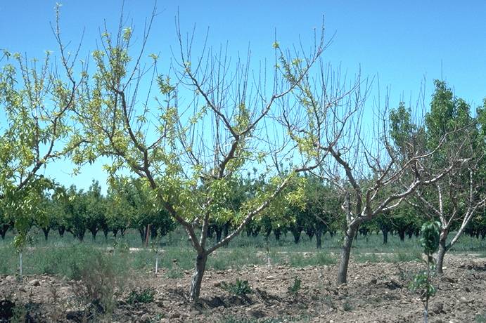
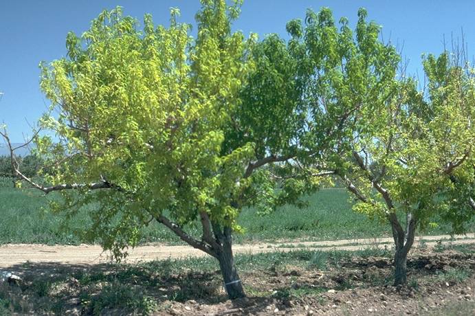
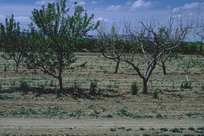
Peach,_chronic_chlorosis_(iron_deficiency),_tree_dieback
Peach, chronic chlorosis (iron deficiency), tree dieback (H. J. Larsen; Colorado State University)
Peach,_Complete_Green,_phytotoxicity
Peach, Complete Green, phytotoxicity (A. R. Renquist; Colorado State University)
Peach,_Complete_Green,_phytotoxicity,_leaf_injury
Peach, Complete Green, phytotoxicity, leaf injury (A. R. Renquist; Colorado State University)
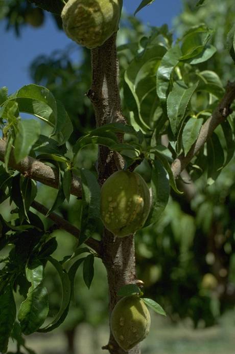
Peach,_Coryneum_blight,_fruit_infection
Peach, Coryneum blight, fruit infection (N. S. Luepschen; Colorado State University)
Peach,_Coryneum_blight,_fruit_infection
Peach, Coryneum blight, fruit infection (N. S. Luepschen; Colorado State University)
Peach,_Coryneum_blight,_fruit_infection
Peach, Coryneum blight, fruit infection (N. S. Luepschen; Colorado State University)
Peach,_Coryneum_blight,_fruit_infection_(late_season)
Peach, Coryneum blight, fruit infection (late season)
(H. J. Larsen; Colorado State University)

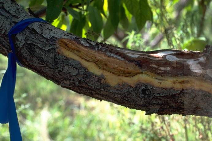
Peach,_Cytospora_canker,_infection_process
Peach, Cytospora canker, infection process (S. Savage; Colorado State University)
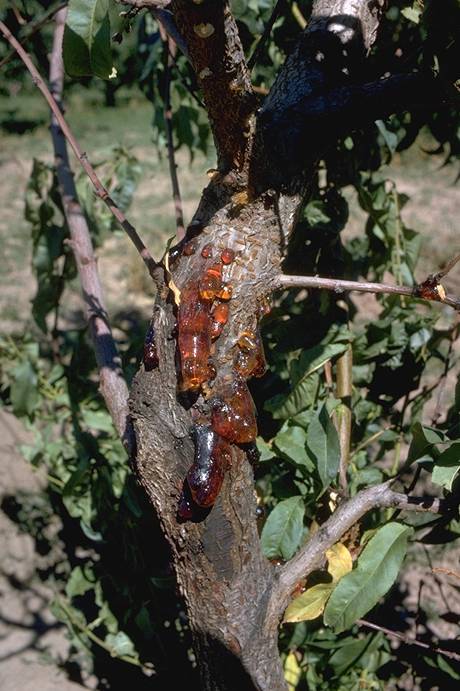
Peach,_Cytospora_canker_(gummosis)_symptoms
Peach, Cytospora canker (gummosis) symptoms (N. S. Luepschen ; Colorado State University)
Peach,_Cytospora_canker_(gummosis)_symptoms
Peach, Cytospora canker (gummosis) symptoms (N. Luepschen; Colorado State University)

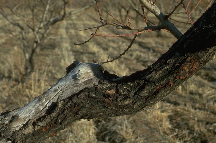
Peach,_Cytospora_canker_+_sunscald
Peach, Cytospora canker + sunscald (N. S. Luepschen; Colorado State University)
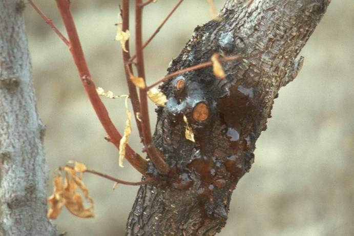
Peach,_Cytospora_canker_infection_at_pruning_cuts
Peach, Cytospora canker infection at pruning cuts (N. S. Luepschen; Colorado State University)
Peach,_freeze_&_winter_injury,_shoots
Peach, freeze & winter injury, shoots (H. J. Larsen; Colorado State University)
Peach,_hail_damage
Peach, hail damage (N. S. Luepschen; Colorado State University)
Peach,_hail_damage,_fruit
Peach, hail damage, fruit (H. J. Larsen; Colorado State University)
Peach,_hail_damage,_tree_bark
Peach, hail damage, tree bark (H. J. Larsen; Colorado State University)
Peach,_hail_damage,_tree_bark
Peach, hail damage, tree bark (H. J. Larsen; Colorado State University)
Peach,_herbicide_damage
Peach, herbicide damage (A. R. Renquist; Colorado State University)
Peach,_herbicide_damage
Peach, herbicide damage (A. R. Renquist; Colorado State University)
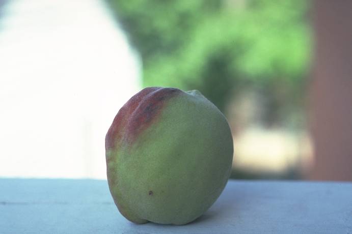
Peach,_inking_(abiotic)_-_severe_on_right
Peach, inking (abiotic) - severe on right (H. J. Larsen; Colorado State University)
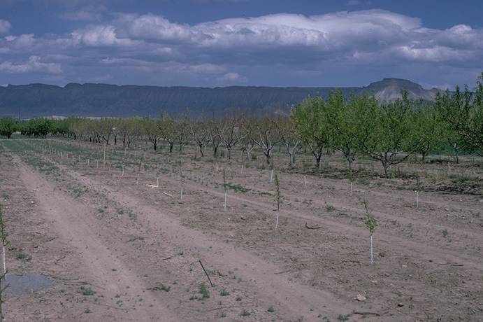
Peach,_iron_deficiency_chlorosis_control_with_iron_chelate
Peach, iron deficiency chlorosis control with iron chelate (C. R. Ure; Colorado State University)
Peach,_Kearneyville_free_Y,_perpendicular_V
Peach, Kearneyville free Y, perpendicular V (A. R Renquist; Colorado State University)
Peach,_leaf_cover_+_herbicide_timing
Peach, leaf cover + herbicide timing (A. R. Renquist; Colorado State University)
Peach,_left_dead,_right_viable_&_unopened
Peach, left dead, right viable & unopened (A. R. Renquist; Colorado State University)
Peach,_peach_twig_borer_damage
Peach, peach twig borer damage (H. J. Larsen; Colorado State University)
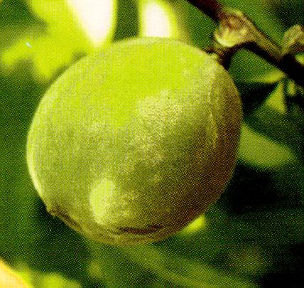
Peach,_powdery_mildew,_fruit_infection_(rusty_spot)_
Peach, powdery mildew, fruit infection (rusty spot) (N. S. Luepschen; Colorado State University)
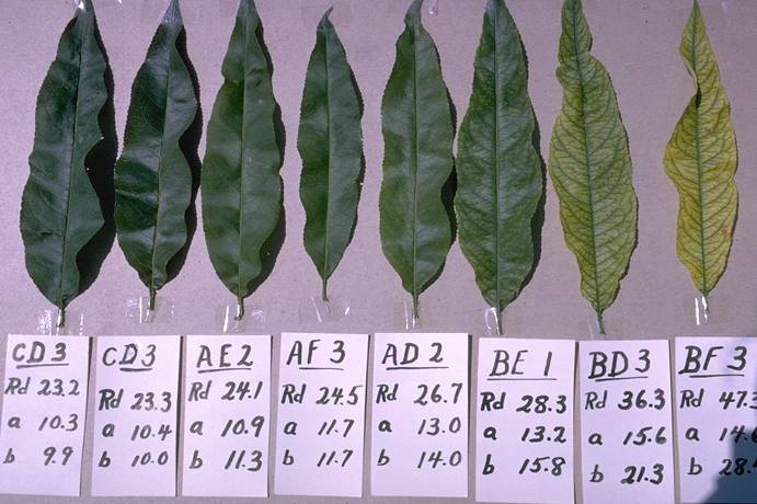
Peach,_range_of_foliage_colors,_winter_injury_recovery_+_fertilizer_trts
Peach, range of foliage colors, winter injury recovery + fertilizer trts (C. R. Ure; Colorado State University)
Peach,_red_suture_(MLO),_fruit_symptoms
Peach, red suture (MLO), fruit symptoms (N. S. Luepschen ; Colorado State University)
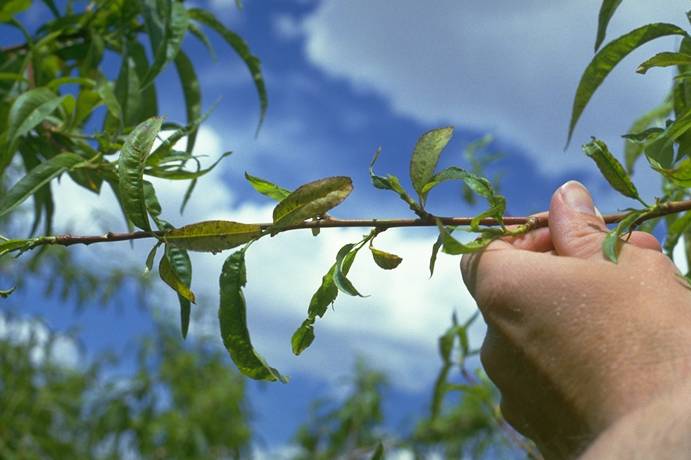
Peach,_red_suture_(MLO),_leaf_&_fruit_symptoms
Peach, red suture (MLO), leaf & fruit symptoms (N. S. Luepschen ; Colorado State University)
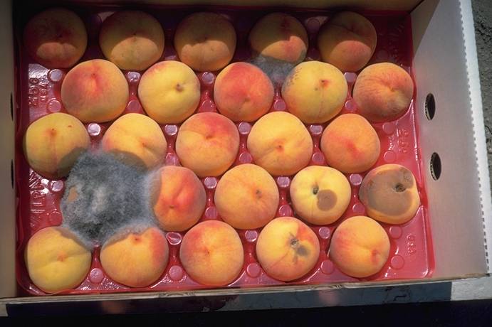
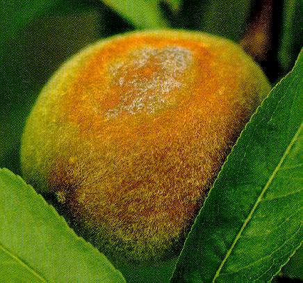
Peach,_Rhizopus,_5_days_post-harvest
Peach, Rhizopus, 5 days post-harvest (N. S. Luepschen; Colorado State University)
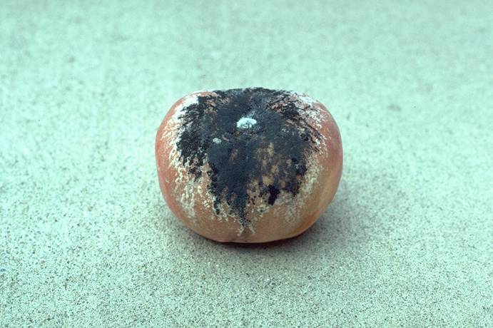
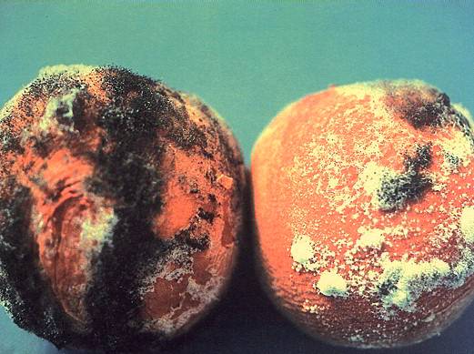
Peach,_Rhizopus_rot_of_fruit
Peach, Rhizopus rot of fruit (H. J. Larsen; Colorado State University)
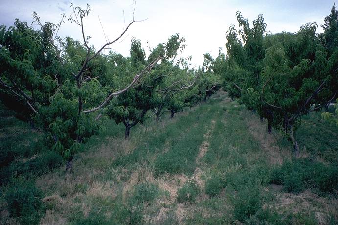
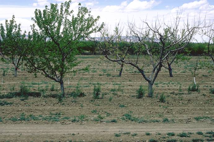
Peach,_severe_chlorosis_(iron_deficiency),_tree_dieback
Peach, severe chlorosis (iron deficiency), tree dieback (H. J. Larsen; Colorado State University)
Peach,_shot-hole_borer_+_woodpecker_feeding
Peach, shot-hole borer + woodpecker feeding (N. S. Luepschen; Colorado State University)
Peach,_snow_damage
Peach, snow damage (H. J. Larsen; Colorado State University)
Peach,_snow_damage
Peach, snow damage (H. J. Larsen; Colorado State University)
Peach,_trunk_cracking/spiral
Peach, trunk cracking/spiral (A. R. Renquist; Colorado State University)
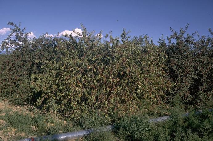
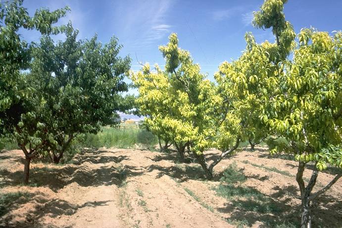
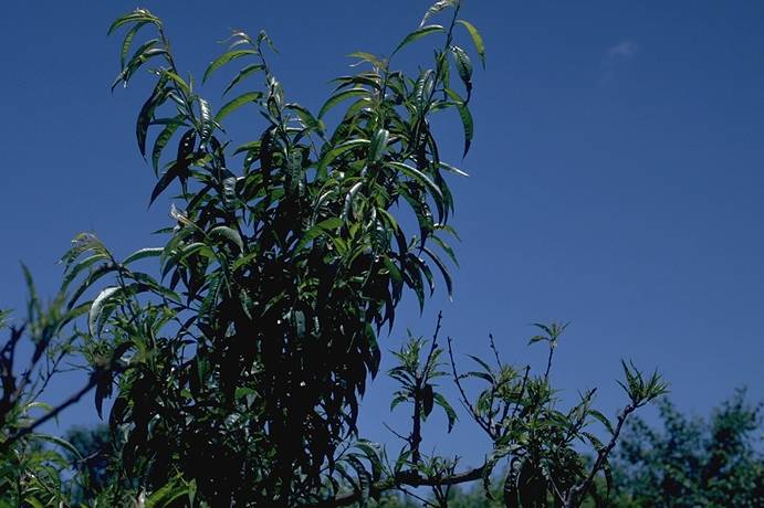
Peach,_western_X_disease,_whole_tree_symptoms
Peach, western X disease, whole tree symptoms (C. L. Parish; Colorado State University)
Peach,_winter_bud_kill
Peach, winter bud kill (H. J. Larsen; Colorado State University)
Peach,_winter_injury,_wood_browning
Peach, winter injury, wood browning (H. J. Larsen; Colorado State University)
Peach,_winter_injury_+_pruning_timing_effects
Peach, winter injury + pruning timing effects (H. J. Larsen; Colorado State University)
Peach,_winter_injury_on_left_(pruned)
Peach, winter injury on left (pruned)
(A. R. Renquist; Colorado State University)
Peach,_X-disease_+_winter_damage
Peach, X-disease + winter damage (H. J. Larsen; Colorado State University)
Peach,_X-disease_+_winter_damage
Peach, X-disease + winter damage (H. J. Larsen; Colorado State University)
Peach,_X-disease_+_winter_damage
Peach, X-disease + winter damage (H. J. Larsen; Colorado State University)
Peach,_X-disease_+_winter_damage
Peach, X-disease + winter damage (H. J. Larsen; Colorado State University)

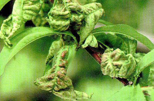
Peach_Leaf_Curl_1
Peach Leaf Curl 1 ==
Deformed peach leaves with leaf curl.
(Alan L. Jones, Michigan State University and Turner B. Sutton, North Carolina State University)
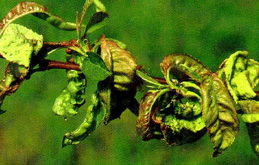
Peach_Leaf_Curl_2
Peach Leaf Curl 2 ==
Peach leaf curl. Note extensive red discoloration compared with leaves shown in Photo 113.
(Alan L. Jones, Michigan State University and Turner B. Sutton, North Carolina State University)
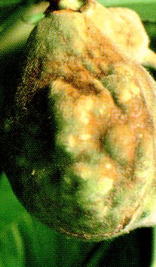
Peach_Leaf_Curl_3a
Peach Leaf Curl 3a ==
Leaf curl on fruit of peach.
(Alan L. Jones, Michigan State University and Turner B. Sutton, North Carolina State University)
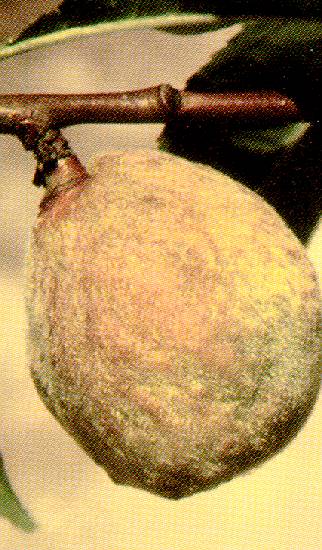
Peach_Leaf_Curl_3b
Peach Leaf Curl 3b ==
Leaf curl on fruit of peach.
(Alan L. Jones, Michigan State University and Turner B. Sutton, North Carolina State University)
Peach_Leaf_Curl_4a
Peach Leaf Curl 4a ==
Asci of the leaf curl fungus emerging from the surface of a leaf.
(Alan L. Jones, Michigan State University and Turner B. Sutton, North Carolina State University)
Peach_Leaf_Curl_4b
Peach Leaf Curl 4b ==
Ruptured ascus containing ascospores and producing a bud conidium.
(Alan L. Jones, Michigan State University and Turner B. Sutton, North Carolina State University)
Peach_Leaf_Curl_4c
Peach Leaf Curl 4c ==
An ascospore producing a bud conidium.
(Alan L. Jones, Michigan State University and Turner B. Sutton, North Carolina State University)
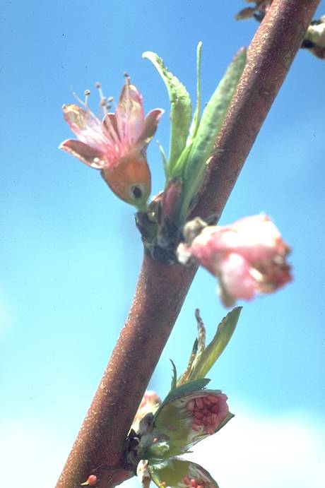
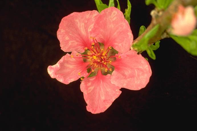
Peach_mosaic,_flower_color_break_symptom
Peach mosaic, flower color break symptom (H. J. Larsen; Colorado State University)
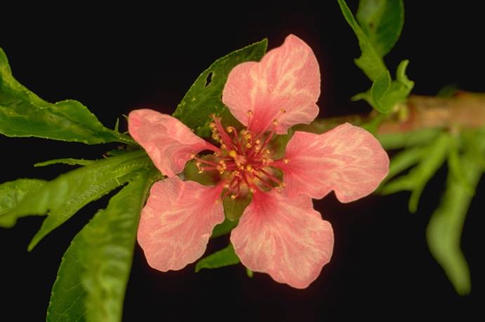
Peach_mosaic,_flower_color_break_symptom
Peach mosaic, flower color break symptom (H. J. Larsen; Colorado State University)
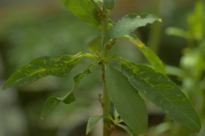
Peach_mosaic,_foliage_symptoms
Peach mosaic, foliage symptoms (H. J. Larsen; Colorado State University)
Peach_mosaic,_foliage_symptoms_(isolate_CL-1)
Peach mosaic, foliage symptoms (isolate CL-1)
(H. J. Larsen; Colorado State University)
Peach_mosaic,_foliage_symptoms_(isolate_CL-2)
Peach mosaic, foliage symptoms (isolate CL-2)
(H. J. Larsen; Colorado State University)
Peach_mosaic,_Loring_peach,_L._healthy,_R._symptomatic_
Peach mosaic, Loring peach, L. healthy, R. symptomatic (H. J. Larsen; Colorado State University)
Peach_mosaic,_peach_&_nectarine_fruit_symptoms
Peach mosaic, peach & nectarine fruit symptoms (H. J. Larsen; Colorado State University)
Peach_mosaic,_vein_clearing_symptoms
Peach mosaic, vein clearing symptoms (N. S. Luepschen; Colorado State University)
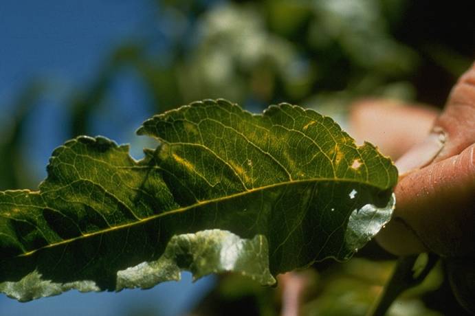
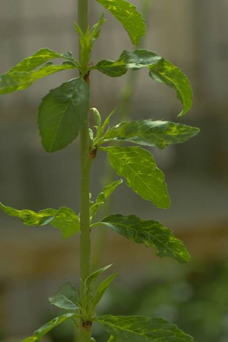
Peach_mosaic_leaf_symptoms,_healthy_vs_infected
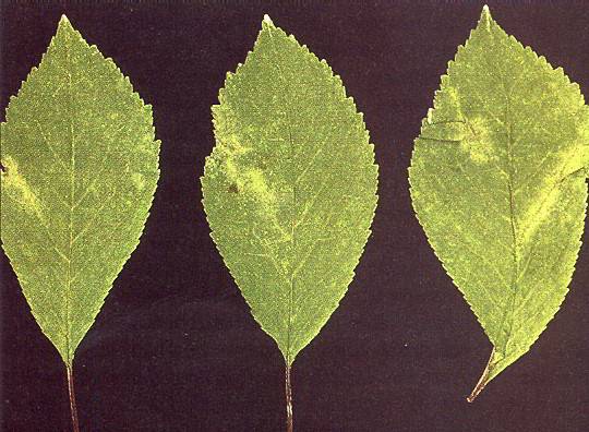
Peach mosaic leaf symptoms, healthy vs infected (H. J. Larsen; Colorado State University)
Peach_Perennial_Canker_1
Peach Perennial Canker 1 ==
Perennial canker of peach.
(Alan L. Jones, Michigan State University and Turner B. Sutton, North Carolina State University)
Peach_Perennial_Canker_2
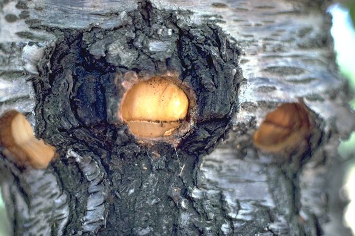
Peach Perennial Canker 2 ==
Peach canker with bark removed to expose black structures with white margins containing the pycnidial stromata of Leucostoma spp.
(Alan L. Jones, Michigan State University and Turner B. Sutton, North Carolina State University)
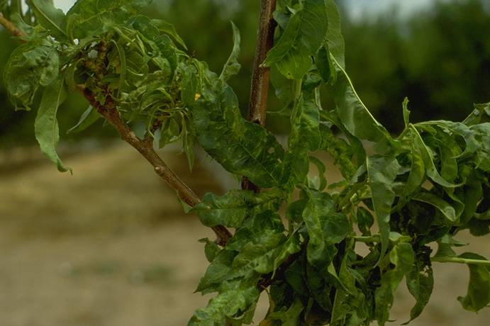
Peach_rosette_MLO
Peach rosette MLO compact cluster on peach leaves (pitfruit, stone fruit, disease) (J.A. Foster, USDA)
Peach_Rosette_Mosaic
Peach Rosette Mosaic ==
Peach tree infected when young with peach rosette mosaic. Note compact growth due to rosetting.
(Alan L. Jones, Michigan State University and Turner B. Sutton, North Carolina State University)

Peach_Scab_1
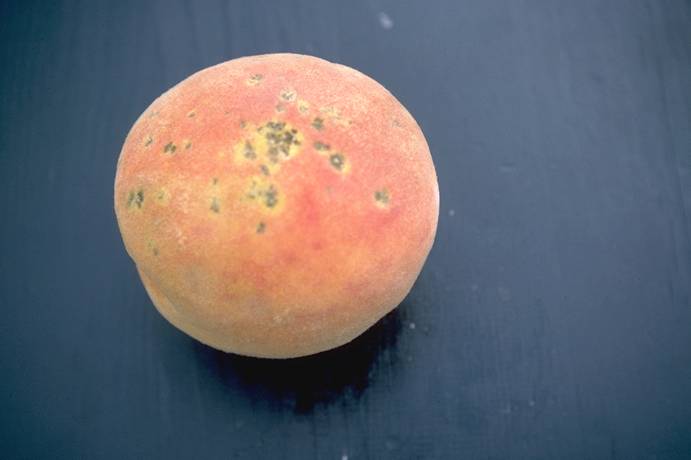
Peach Scab 1 ==
Peach with brown to black, velvety lesions caused by the scab fungus Cladosporium carpophilum.
(Alan L. Jones, Michigan State University and Turner B. Sutton, North Carolina State University)
Peach_Scab_2
Peach Scab 2 ==
Mature peach with brown to black scab lesions with yellow margins.
(Alan L. Jones, Michigan State University and Turner B. Sutton, North Carolina State University)
Peach_Scab_3
Peach Scab 3 ==
Black, depressed lesions on a peach twig caused by the scab fungus Cladosporium carpophilum.
(Alan L. Jones, Michigan State University and Turner B. Sutton, North Carolina State University)
Peach_winter_damage,_pruned_before_2/6/89
Peach winter damage, pruned before 2/6/89 (H. J. Larsen; Colorado State University)
Peach_yellow_bud_strain_of_tomato_ringspot_virus
Peach yellow bud mosaic strain of tomato ringspot virus on peach leaf (pitfruit, stone fruit, disease) (J.K. Uyemoto, USDA)
Peach_yellows_MLO
Peach yellows MLO symptoms: proliferation, upright, slender shoots, small chlorotic leaves, scaffold branch, tree infected, (pitfruit, stone fruit, disease) (J.K. Uyemoto, USDA)
Phytophthora_Root,_Crown,_and_Colar_Rot_1
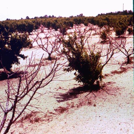
Phytophthora Root, Crown, and Colar Rot 1 ==
Progressive decline and eventual death of Montmorency sour cherry trees from Phytophthora root and crown rot.
(Alan L. Jones, Michigan State University and Turner B. Sutton, North Carolina State University)
Phytophthora_Root,_Crown,_and_Colar_Rot_2
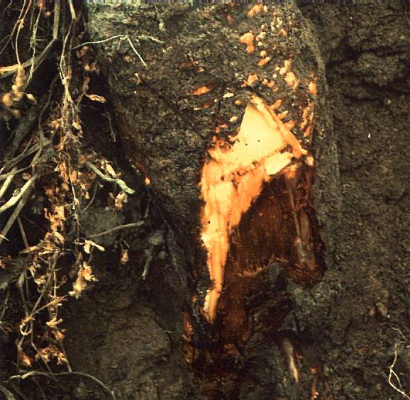
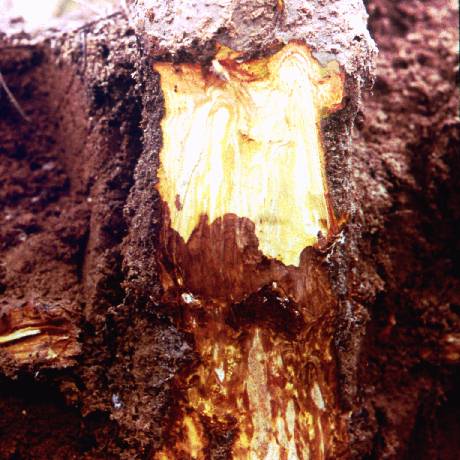
Phytophthora Root, Crown, and Colar Rot 2 ==
Decay of Mahaleb rootstock below ground caused by Phytophthora root and crown rot.
(Alan L. Jones, Michigan State University and Turner B. Sutton, North Carolina State University)
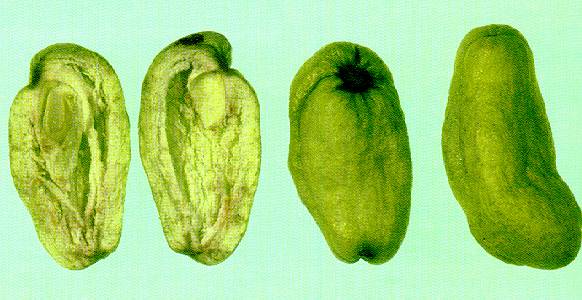
Plum_Pockets_1
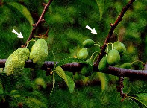
Plum Pockets 1 ==
Plum pockets. Note enlarged infected fruit on left and normal-sized healthy fruit on right.
(Alan L. Jones, Michigan State University and Turner B. Sutton, North Carolina State University)
Plum_Pockets_2
Plum Pockets 2 ==
Hollow interior of plum pockets-infected plums.
(Alan L. Jones, Michigan State University and Turner B. Sutton, North Carolina State University)
Plum_pox_virus
Plum pox virus fruit symptoms on apricot (pitfruit, stone fruit, disease) (J.A. Foster, USDA)
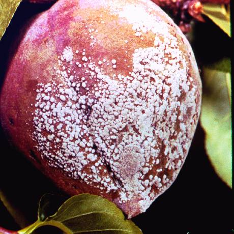
Powdery_Mildew/Rusty_Spot_of_Peach_1
Powdery Mildew/Rusty Spot of Peach 1 ==
White fungal growth of the powdery mildew fungus on peach.
(Alan L. Jones, Michigan State University and Turner B. Sutton, North Carolina State University)
Powdery_Mildew/Rusty_Spot_of_Peach_2a
Powdery Mildew/Rusty Spot of Peach 2a ==
Reddish (left) lesions on peach associated with powdery mildew.
(Alan L. Jones, Michigan State University and Turner B. Sutton, North Carolina State University)
Powdery_Mildew/Rusty_Spot_of_Peach_2b
Powdery Mildew/Rusty Spot of Peach 2b ==
Rusty-colored (right) lesions on peach associated with powdery mildew.
(Alan L. Jones, Michigan State University and Turner B. Sutton, North Carolina State University)
Powdery_Mildew_of_Cherry_1
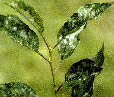
Powdery Mildew of Cherry 1 ==
White fungal growth of the powdery mildew fungus on leaves of sour cherry.
(Alan L. Jones, Michigan State University and Turner B. Sutton, North Carolina State University)
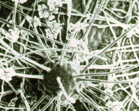
Powdery_Mildew_of_Cherry_2
Powdery Mildew of Cherry 2 ==
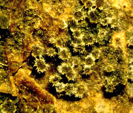
Cleistothecium of cherry mildew with dichotomous branching of appendages.
(Alan L. Jones, Michigan State University and Turner B. Sutton, North Carolina State University)
Powdery_Mildew_of_Cherry_3
Powdery Mildew of Cherry 3 ==
Red blotches on Montmorency sour cherry caused by powdery mildew.
(Alan L. Jones, Michigan State University and Turner B. Sutton, North Carolina State University)
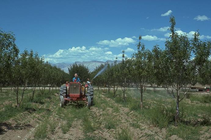
Powdery_mildew_spraying_(handgun_application)
Powdery mildew spraying (handgun application)
(N. S. Luepschen; Colorado State University)
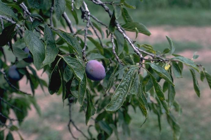
Prune_dwarf_virus_symptoms_on_Italian_prune
Prune dwarf virus symptoms on Italian prune (H. J. Larsen; Colorado State University)
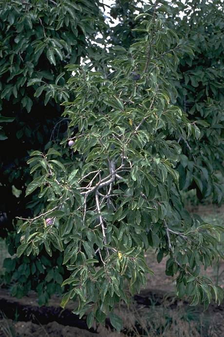
Prune_dwarf_virus_symptoms_on_Italian_prune
Prune dwarf virus symptoms on Italian prune (H. J. Larsen; Colorado State University)
Prunus_necrotic_ringspot_ilarvirus
Prunus necrotic ringspot ilarvirus vein banding symptoms on kelsey plum (pitfruit, stone fruit, disease) (J.K. Uyemoto, USDA)

Prunus_Necrotic_Ringspot_1
Prunus Necrotic Ringspot 1 ==
Depressed, concentric rings on the upper surface of a sour cherry leaf due to prunus necrotic ringspot virus.
(Alan L. Jones, Michigan State University and Turner B. Sutton, North Carolina State University)

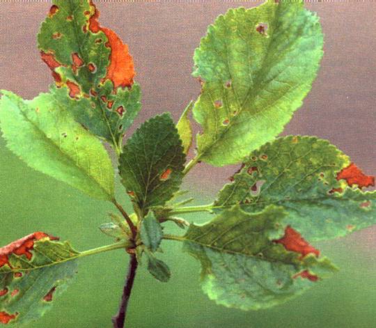
Prunus_Necrotic_Ringspot_2
Prunus Necrotic Ringspot 2 ==
Pattern of shock symptoms on spur leaves of sour cherry caused by Prunus necrotic ringspot virus.
(Alan L. Jones, Michigan State University and Turner B. Sutton, North Carolina State University)
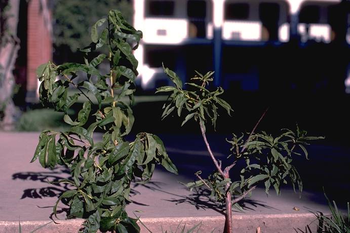
Prunus_Stem_Pitting/Prune_Brownline_1
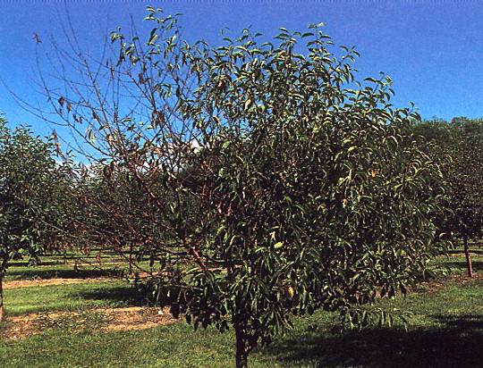
Prunus Stem Pitting/Prune Brownline 1 ==
Peach tree in decline and healthy tree of the same age.
(Alan L. Jones, Michigan State University and Turner B. Sutton, North Carolina State University)
Prunus_Stem_Pitting/Prune_Brownline_2
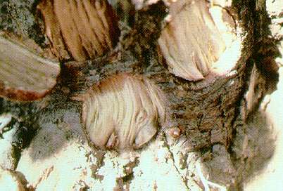
Prunus Stem Pitting/Prune Brownline 2 ==
Sour cherry tree with bark removed to show pitting in the Mahaleb rootstock caused by Prunus stem pitting.
(Alan L. Jones, Michigan State University and Turner B. Sutton, North Carolina State University)
Prunus_Stem_Pitting/Prune_Brownline_3a
Prunus Stem Pitting/Prune Brownline 3a ==
Prunus stem pitting on Stanley plum. Note constriction of Myrobalan rootstock and shoots from the trunk that indicate the constriction.
(Alan L. Jones, Michigan State University and Turner B. Sutton, North Carolina State University)
Prunus_Stem_Pitting/Prune_Brownline_3b
Prunus Stem Pitting/Prune Brownline 3b ==
Prunus stem pitting on Stanley plum. Note constriction of Myrobalan rootstock and shoots from the trunk that indicate the constriction.
(Alan L. Jones, Michigan State University and Turner B. Sutton, North Carolina State University)
Prunus_Stem_Pitting/Prune_Brownline_4
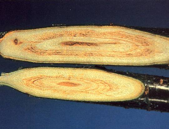
Prunus Stem Pitting/Prune Brownline 4 ==
Prunus stem pitting. Bark was removed to show wide line of dead tissue at the union between the Stanley plum scion and the Myrobalan rootstock.
(Alan L. Jones, Michigan State University and Turner B. Sutton, North Carolina State University)
Rhizopus
Rhizopus ==
Rhizopus rot on peach.
(Alan L. Jones, Michigan State University and Turner B. Sutton, North Carolina State University)
Root_Rot
Root Rot
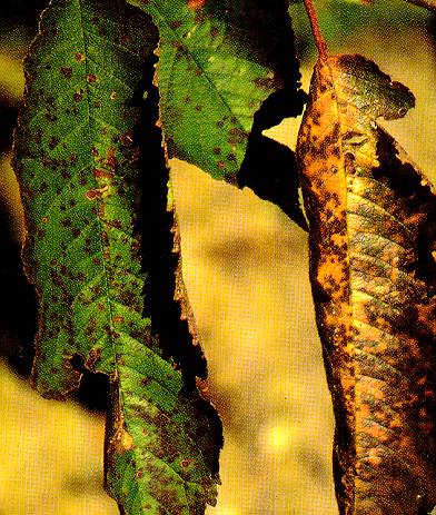
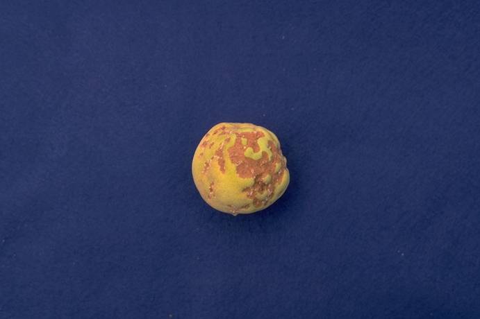
Rusty_blotch
Rusty blotch leaf symptoms on santa rosa plum: chlorosis along leaf margins (healthy leaf, lower right) (pitfruit, stone fruit, disease) (J.K. Uyemoto, USDA)
Scab_on_Fruit
Scab on Fruit
Scab_on_Twig
Scab on Twig

Silver_Leaf
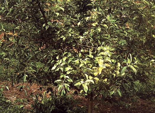
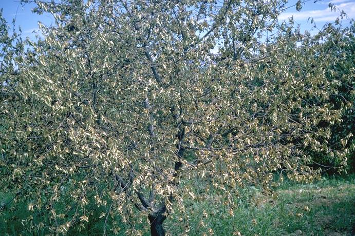
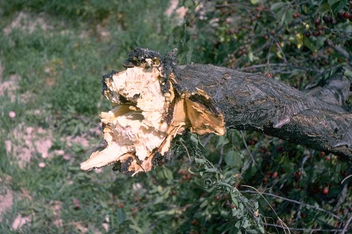
Silver Leaf ==
Silver leaf symptoms on apple leaves. Symptoms on cherries are similar.
(Alan L. Jones, Michigan State University and Turner B. Sutton, North Carolina State University)
Sour_cherry,_crown_borer_damage
Sour cherry, crown borer damage (N. S. Luepschen; Colorado State University)

Sour_cherry,_powdery_mildew,_leaf_infection
Sour cherry, powdery mildew, leaf infection (N. S. Luepschen; Colorado State University)

Sour_cherry,_snow_damage
Sour cherry, snow damage (H. J. Larsen; Colorado State University)

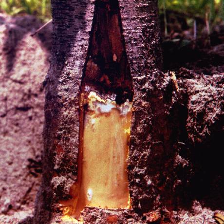
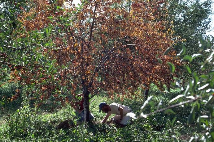
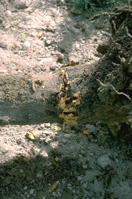
Sour_cherry_decline,_crown_borer
Sour cherry decline, crown borer (N. S. Luepschen; Colorado State University)
Sour_cherry_decline,_crown_borer
Sour cherry decline, crown borer (N. S. Luepschen; Colorado State University)
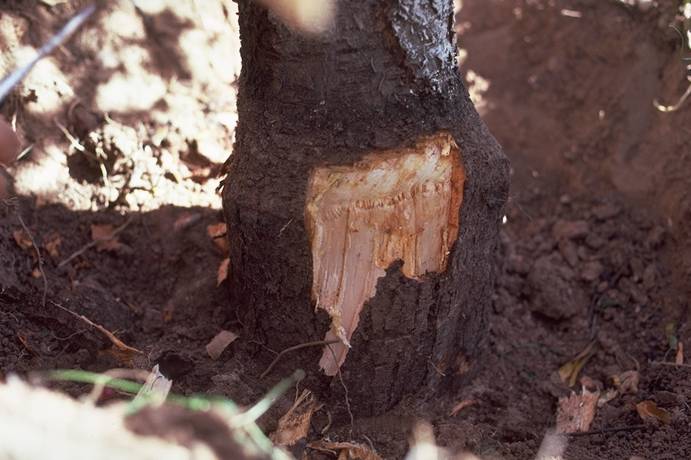
Sour_cherry_decline,_crown_borer_injury
Sour cherry decline, crown borer injury (N. S. Luepschen; Colorado State University)
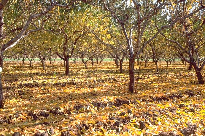
Sour_Cherry_Yellows
Sour Cherry Yellows ==
Yellowing and defoliation of sour cherry leaves due to sour cherry yellows.
(Alan L. Jones, Michigan State University and Turner B. Sutton, North Carolina State University)
Sweet_cherry,_cherry_rasp-leaf_virus,_symptoms
Sweet cherry, cherry rasp-leaf virus, symptoms (N. S. Luepschen; Colorado State University)
Sweet_cherry,_Coryneum_leaf_infection
Sweet cherry, Coryneum leaf infection (N. S. Luepschen; Colorado State University)
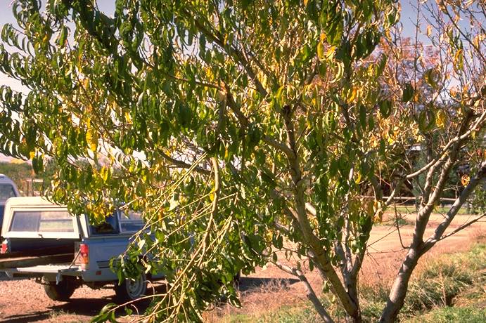
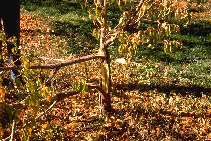
Sweet_cherry,_Cytospora_collapse
Sweet cherry, Cytospora collapse (H. J. Larsen; Colorado State University)
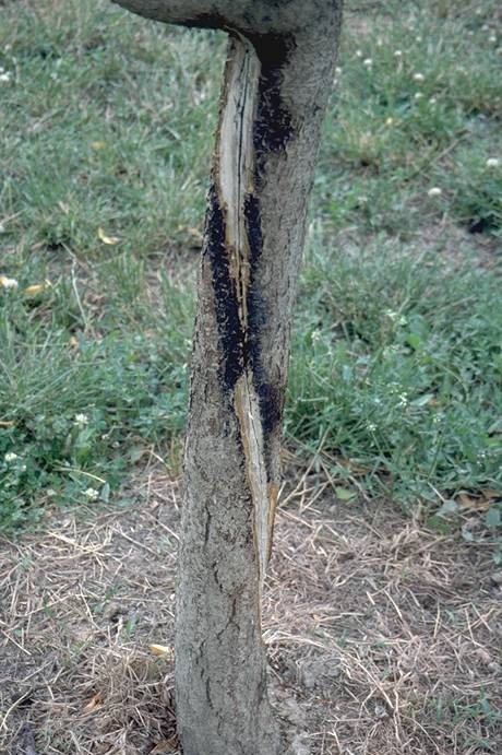
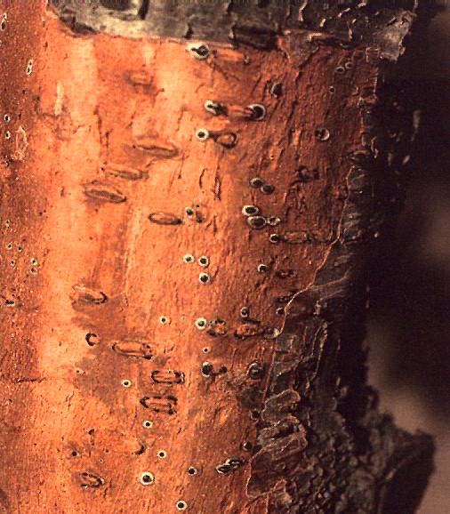
Sweet_cherry,_Cytospora_fruiting_bodies_beneath_bark_epidermis
Sweet cherry, Cytospora fruiting bodies beneath bark epidermis (H. J. Larsen; Colorado State University)
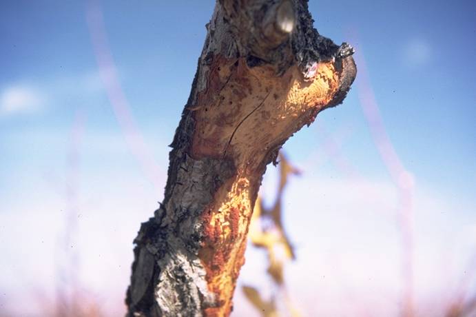
Sweet_cherry,_Cytospora_infection_progress
Sweet cherry, Cytospora infection progress (H. J. Larsen; Colorado State University)
Sweet_cherry,_powdery_mildew_on_leaf_(healthy_left)
Sweet cherry, powdery mildew on leaf (healthy left)
(N. S. Luepschen; Colorado State University)
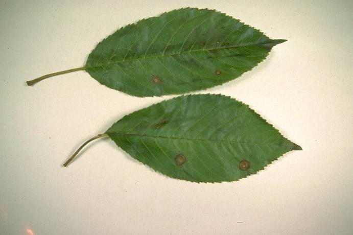
Sweet_cherry,_Prunus_rusty_mottle_virus,_leaf_symptoms
Sweet cherry, Prunus rusty mottle virus, leaf symptoms (H. J. Larsen; Colorado State University)
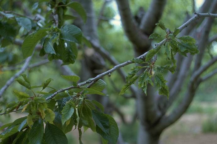
Sweet_cherry,_Prunus_rusty_mottle_virus,_tree_symptoms
Sweet cherry, Prunus rusty mottle virus, tree symptoms (H. J. Larsen; Colorado State University)
Sweet_cherry,_Verticillium_wilt,_whole_tree_symptoms
Sweet cherry, Verticillium wilt, whole tree symptoms (H. J. Larsen; Colorado State University)
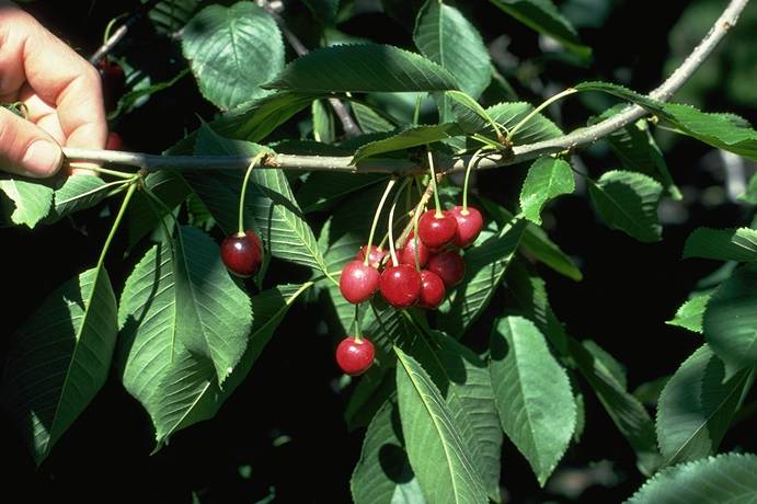
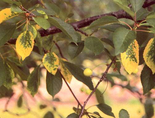
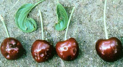
Sweet_cherry,_X-disease,_fruit_symptoms
Sweet cherry, X-disease, fruit symptoms (C. L. Parish; Colorado State University)
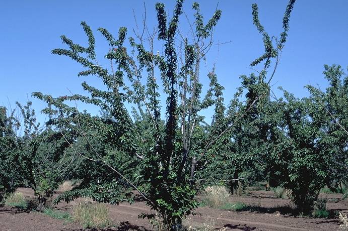
Sweet_cherry,_X-disease_(MLO),_tree_decline_(Mazzard_rootstock)
Sweet cherry, X-disease (MLO), tree decline (Mazzard rootstock)
(H. J. Larsen; Colorado State University)
Sweet_cherry_&_peach,_cherry_rasp-leaf,_symptoms_on_leaves
Sweet cherry & peach, cherry rasp-leaf, symptoms on leaves (N. S. Luepschen; Colorado State University)

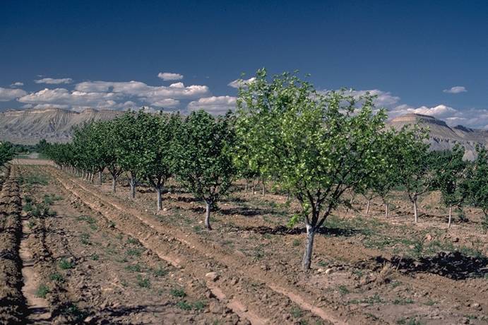
Tart_cherry,_X-disease,_tree_collapse_(Mahaleb_rootstock)
Tart cherry, X-disease, tree collapse (Mahaleb rootstock)
(H. J. Larsen; Colorado State University)
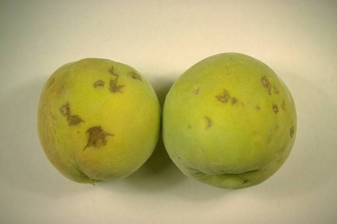
Tomato_bushy_stunt_virus
Tomato bushy stunt virus on sam sweet cherry: deformed leaves, necrotic fruit symptoms (healthy fruit, right) (pitfruit, stone fruit, disease) (J.K. Uyemoto, USDA)
Tomato_ringspot_virus
Peach yellow bud mosaic strain of tomato ringspot virus: small, yellow leaf clusters (lower vegetative buds), normal leaf cluster (far right, upper bud), larger, green (pitfruit, stone fruit, disease) (J.K. Uyemoto, USDA)

Tomato_ringspot_virus
Tomato ringspot virus on president plum with peach rootstock (brown line partially formed along, scion-rootstock junction) (pitfruit, stone fruit, disease) (J.K. Uyemoto, USDA)
Tomato_ringspot_virus
Tomato ringspot virus symptoms on peach rootstock: stem pitting, woody cylinder (pitfruit, stone fruit, disease) (Uyemoto, USDA)
Verticillium_Wilt_1
Verticillium Wilt 1 ==
Defoliation and wilting of lateral branches of Montmorency sour cherry caused by Verticillium wilt.
(Alan L. Jones, Michigan State University and Turner B. Sutton, North Carolina State University)
Verticillium_Wilt_2
Verticillium Wilt 2 ==
Grayish streaks in the sapwood of Montmorency sour cherry with Verticillium wilt.
(Alan L. Jones, Michigan State University and Turner B. Sutton, North Carolina State University)
X-Disease_1
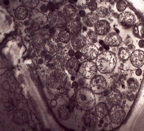
X-Disease 1 ==
Cross-section of a vein in a peach leaf infected with X-disease. The spherical bodies are phytoplasmas and are found in a phloem cell.
(Alan L. Jones, Michigan State University and Turner B. Sutton, North Carolina State University)
X-Disease_2
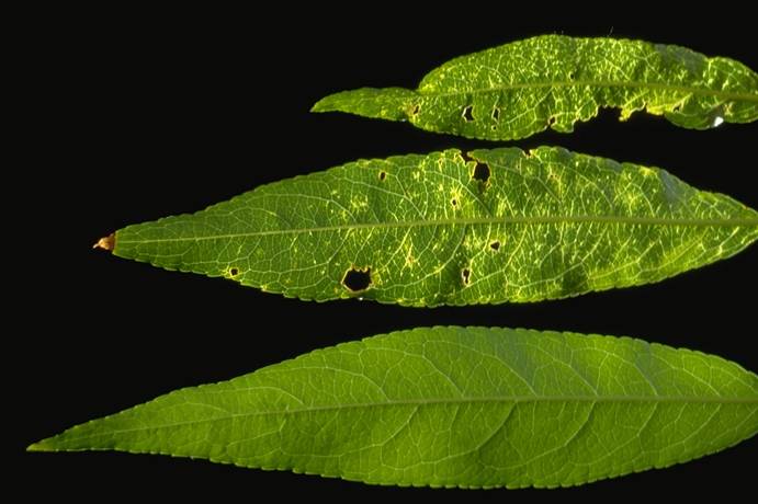
X-Disease 2 ==
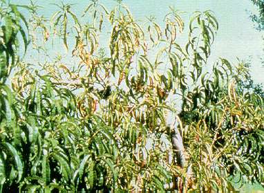
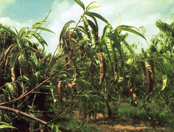
Peach leaves with rolling, red blotch and tattering due to X-disease. Note defoliation starting at the base of the shoot.
(Alan L. Jones, Michigan State University and Turner B. Sutton, North Carolina State University)
X-Disease_3
X-Disease 3 ==
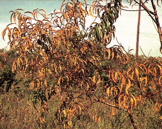
Peach leaves yellowed and rolled from X-disease in late August.
(Alan L. Jones, Michigan State University and Turner B. Sutton, North Carolina State University)
X-Disease_4
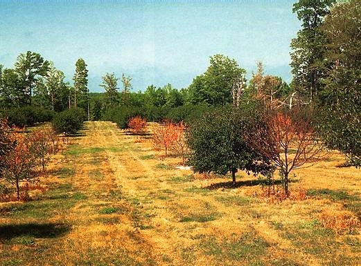
X-Disease 4 ==
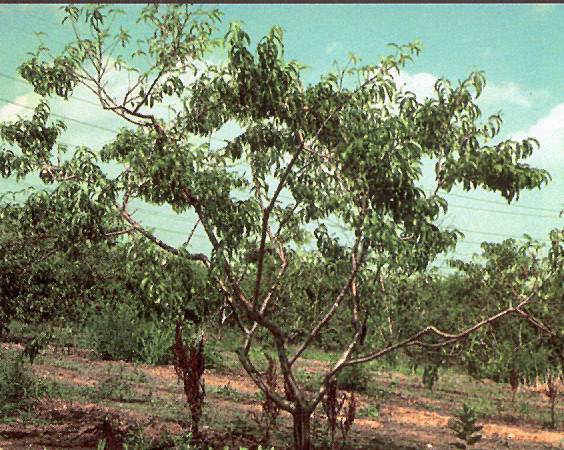
Symptoms of X-disease randomly distributed throughout tree.
(Alan L. Jones, Michigan State University and Turner B. Sutton, North Carolina State University)
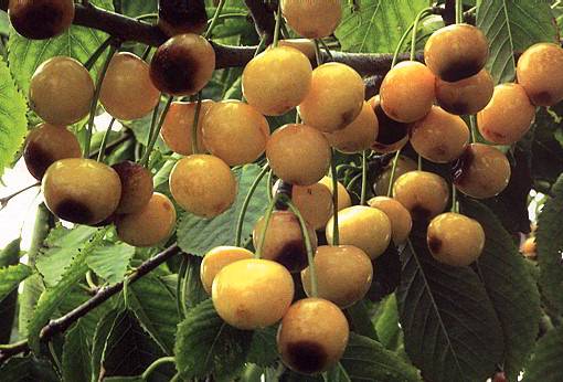
X-Disease_5
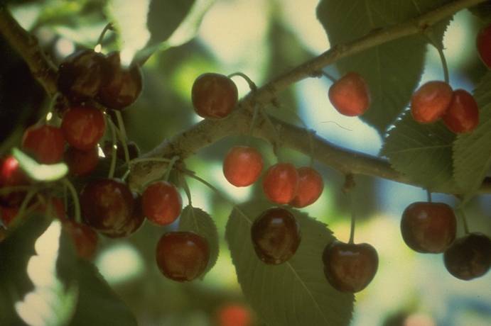
X-Disease 5 ==
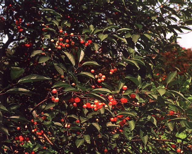
Green to half-ripe cherries interspersed with normal fruit on a sour cherry tree on Mazzard rootstock infected with X-disease.
(Alan L. Jones, Michigan State University and Turner B. Sutton, North Carolina State University)
X-Disease_6
X-Disease 6 ==
Sweet cherry leaf with enlarged stipules due to X-disease.
(Alan L. Jones, Michigan State University and Turner B. Sutton, North Carolina State University)
X-Disease_7
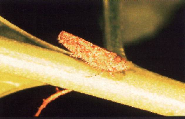
X-Disease 7 ==
Paraphlepsius irroratus, one of several species of leafhoppers that can spread X-disease.
(Alan L. Jones, Michigan State University and Turner B. Sutton, North Carolina State University)