Apples Pears:Photos
Apple and Pear Pest and Nutritional Disorder Photos
Contents
- 1 2,4-D_herbicide_injury_on_pear
- 2 Alternaria_Blotch_1
- 3 Alternaria_Blotch_2
- 4 Apple_bitter_pit,_calcium_deficiency
- 5 Apple_crown_gall
- 6 Apple_crown_gall
- 7 Apple_maggot_damage,_external_appearance_of_fruit
- 8 Apple_maggot_damage,_internal_&_external_appearance_of_fruit
- 9 Apple_maggot_fruit_damage
- 10 Apple_Mosaic
- 11 Apple_mosaic_virus,_leaf_symptoms_on_Golden_Delicious
- 12 Apple_replant_disease_(ARD),_response_to_fall_preplant_fumigation
- 13 Apple_replant_problem,_left_non-fumigated,_right_fumigated
- 14 Apple_replant_study_-_ARD_bioassay_results_@_76_days
- 15 Apple_scab_fruit_infection
- 16 Apple_scab_lesion_1
- 17 Apple_scab_lesion_2
- 18 Apple_scab_lesion_3
- 19 Apple_scab_lesion_4
- 20 Apple_scab_lesion_5
- 21 Apple_scab_lesion_6
- 22 Apple_scab_lesion_7
- 23 Apple_scab_lesion_8
- 24 Apple_scab_lesion_9
- 25 Apple_Union_Necrosis_and_Decline_1
- 26 Apple_Union_Necrosis_and_Decline_2
- 27 Apple_Union_Necrosis_and_Decline_3
- 28 Apple_winter_injury
- 29 ARD_symptoms_of_poor_growth_on_2nd_leaf_tree,_non-fumigated
- 30 Bitter_Rot_
- 31 Bitter_Rot_1
- 32 Bitter_Rot_2
- 33 Bitter_Rot_3
- 34 Bitter_Rot_4
- 35 Bitter_Rot_5
- 36 Black_Pox
- 37 Black_Root_Rot
- 38 Black_Rot_
- 39 Black_Rot_1
- 40 Black_Rot_2
- 41 Black_Rot_Mummy_and_Frog-Eye_Leaf_Spot_
- 42 Blister_bark_3
- 43 Blister_Spot_1
- 44 Blister_Spot_2
- 45 Blister_Spot_3
- 46 Blossom_Blast_of_Pear
- 47 Blotch
- 48 Brooks_Spot_1
- 49 Brooks_Spot_2
- 50 Brooks_Spot_3
- 51 Bull's-eye_Rot
- 52 Calcium_deficiency,_Jonathan_spot
- 53 Calcium_deficiency_corky_spot_on_d'_Anjou_pear
- 54 Cedar_Apple_Rust_Gall
- 55 Cedar_Apple_Rust_on_Leaf_
- 56 Cherry_raspleaf_virus,_normal_vs_symptomatic_fruit
- 57 Concentric_ring_pattern_1
- 58 Cycle_apple_scab
- 59 Cycle_blight
- 60 Cycle_powdery_mildew
- 61 Cycle_rust
- 62 Cytospora_canker_dieback_on appple
- 63 Cytospora_canker_infection_growth_on_apple
- 64 Cytospora_canker_lesions_on_apple
- 65 Cytospora_canker_of_apple
- 66 Cytospora_spore_threads_on_apple
- 67 Dry-Eye_and_Calyx-end_Rots_1
- 68 Dry-Eye_and_Calyx-end_Rots_2
- 69 European_canker_(Nectria_galligena)_fruiting_bodies_on_apples
- 70 European_canker_on_apple
- 71 Fabraea_Leaf_Spot_1
- 72 Fabraea_Leaf_Spot_2
- 73 Fabraea_Leaf_Spot_3
- 74 Fire_Blight
- 75 Fire_blight_1
- 76 Fire_blight_2
- 77 Fire_blight_3
- 78 Fire_blight_4
- 79 Fire_blight_5
- 80 Fire_blight_6
- 81 Fire_blight_7
- 82 Fire_blight_8
- 83 Fire_blight_canker_on_pear
- 84 Fire_blight_canker_with_ooze_on_pear
- 85 Fire_blight_infection_&_scaffold_loss_of_apple
- 86 Fire_blight_on_2bartlett_pear_
- 87 Fire_blight_on_pear_shoots
- 88 Flat_apple_(cherry_raspleaf_virus)_tree_symptoms_(growth_pattern)
- 89 Freeze_damage_to_apple_flowers
- 90 Freeze_damage_to_apple_flowers
- 91 Freeze_damage_to_bartlett_pear_shoots
- 92 Freeze_injury_to_apple_shoots
- 93 Frost_injury/hail_injury
- 94 Golden_apple,_northwestern_anthracnose_canker
- 95 Golden_Delicious_apple,_apple_wood_rot,_(Trametes_hispida)
- 96 Golden_Delicious_apple,_apple_wood_rot_(Trametes_hispida)
- 97 Golden_Delicious_apple_frost_injury
- 98 Golden_Delicious_apple_iron_chlorosis_(deficiency)_leaf_symptoms
- 99 Gray_Mold
- 100 Hail_damage_on_pear_tree_bark
- 101 Hail_damage_to_apple_fruit
- 102 Jonathan_apple_slime_flux_kill,_cambium_injury_symptoms
- 103 Jonathan_apple_slime_flux_on_lower_trunk
- 104 Jonathan_spot,_internal_calcium_deficiency
- 105 Leucostoma_Canker_1
- 106 Leucostoma_Canker_2
- 107 Moldy_Core_1
- 108 Moldy_Core_2
- 109 Mycosphaerella_Leaf_Spot
- 110 Necrotic_Leaf_Blotch_1
- 111 Necrotic_Leaf_Blotch_2
- 112 Nectria_Canker
- 113 Nectria_Twig_Blight_1
- 114 Nectria_Twig_Blight_2a
- 115 Nectria_Twig_Blight_2b
- 116 Nectria_Twig_Blight_3
- 117 Northwestern_anthracnose_canker_on_apple
- 118 Orchard,_overhead_sprinkler_ice_damage
- 119 Pear_decline,_early_fall_leaf_coloration
- 120 Pear_decline_(MLO)_branch_&_foliage_symptoms
- 121 Pear_decline_(MLO)_branch_symptoms
- 122 Pear_decline_(MLO)_growth_symptoms
- 123 Pear_decline_(MLO)_whole_tree_symptoms
- 124 Pear_fire_blight_canker_with_ooze
- 125 Pear_fire_blight_caused_by_hail_damage
- 126 Pear_fire_blight_shoot_infection_+_ooze
- 127 Pear_fire_blight_shoot_infection_symptoms
- 128 Pear_fire_blight_strikes_in_top_of_tree
- 129 Pear_frost_damage
- 130 Pear_frost_injury
- 131 Pear_root_stock_union
- 132 Pear_scab
- 133 Pear_Scab_1
- 134 Pear_Scab_2
- 135 Penicillium_expansum_(blue_mold)_rot_on_pear_fruit
- 136 Penicillium_expansum_(blue_mold)_rot_on_pear_fruit
- 137 Perennial_canker_+_woolly_aphids_on_apple
- 138 Perennial_canker_of_apple
- 139 Phytophthora_collar_rot_symptoms
- 140 Phytophthora_crown_rot,_Early_Jonathan_trunk_canker
- 141 Phytophthora_crown_rot,_Erly_Jonathan_weak_tree_symptoms
- 142 Phytophthora_crown_rot,_premature_fall_leaf_coloration
- 143 Phytophthora_Root,_Crown,_and_Collar_Rot
- 144 Phytophthora_root_rot_symptoms_of_Jonathan_apple
- 145 Powdery_Mildew
- 146 Powdery_Mildew_1
- 147 Powdery_Mildew_2
- 148 Powdery_Mildew_3
- 149 Powdery_mildew_apple_shoot_infection
- 150 Powdery_mildew_on_apple_2nd_growth_flush
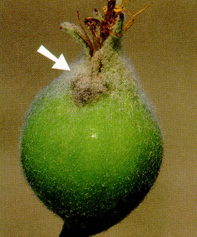
- 151 Powdery_mildew_pear_fruit_infection
- 152 Powdery_mildew_pear_shoot_infection
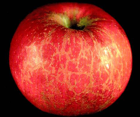
- 153 Powdery_mildew_russet_on_Jonathan_apple_fruit_(right_-_healthy)
- 154 Powdery_mildew_vs_non_mildewed_apple_trees
- 155 Red_delicious_apple,_blister_bark_(virus)_symptoms
- 156 Red_delicious_apple,_dapple_apple_(viroid)_fruit_symptoms
- 157 Red_delicious_apple,_flat_apple_(cherry_raspleaf_virus)_fruit_symptoms
- 158 Rome_apple_blue_mold
- 159 Rome_apple_fire_blight_infection_on_scaffold
- 160 Rome_apple_sunscald_+_bridge_graft
- 161 Rust_1
- 162 Rust_2
- 163 Rust_3
- 164 Rust_4
- 165 Rust_5
- 166 Rust_6
- 167 Rust_7
- 168 Rust_8a
- 169 Rust_8b
- 170 Scab_on_Fruit_
- 171 Scab_on_Young_Fruit_and_Leaf
- 172 Slime_flux_on_black_Jonathan_apple
- 173 Soft_Rot/Blue_Mold_1
- 174 Soft_Rot/Blue_Mold_2
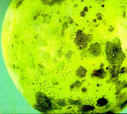
- 175 Sooty_Bloch_and_Flyspeck_1
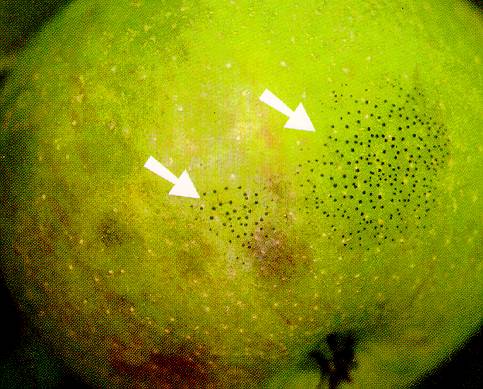
- 176 Sooty_Bloch_and_Flyspeck_2
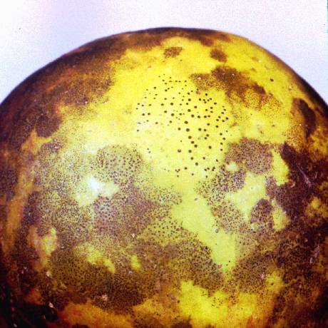
- 177 Sooty_Blotch_and_Flyspeck
- 178 Southern_Blight_1
- 179 Southern_Blight_2
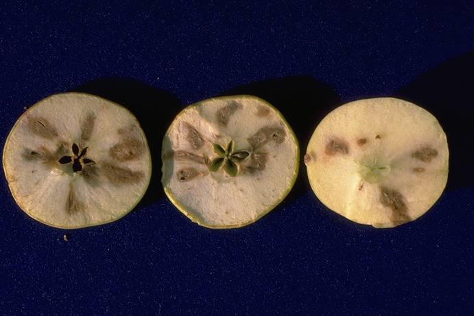
- 180 Stony_Pit_of_Pear
- 181 Sulfur_burn_to_apple_foliage
- 182 Sulfur_burn_to_apple_foliage
- 183 Sulfur_burn_to_apple_fruit_and_foliage
- 184 Thread_Blight
- 185 Union_necrosis_(TmRS_virus)_of_apple
- 186 White_Root_Rot
- 187 White_Rot_1
- 188 White_Rot_2
- 189 White_Rot_3
- 190 White_Rot_4
- 191 White_Rot_5
- 192 Woolly_aphids_on_apple
- 193 X_disease_vector_adult,_Fieberiella_florii
- 194 X_disease_vector_adult,_Fieberiella_florii
- 195 X_disease_vector_adult,_Scaphytopius_acutus
- 196 X_disease_vector_adult_&_nymph,_Fieberiella_florii
- 197 X-Spot
- 198 Zinc_deficiency_1
- 199 Zinc_deficiency_2
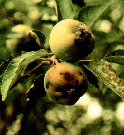
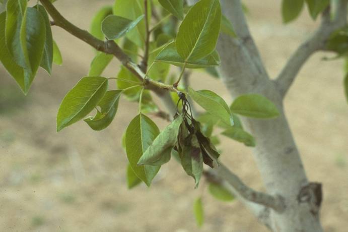
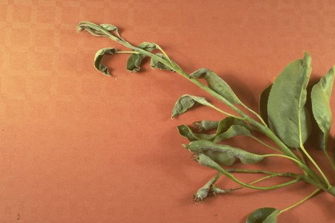
2,4-D_herbicide_injury_on_pear
2,4-D herbicide injury on pear (N. S. Luepschen; Colorado State University)
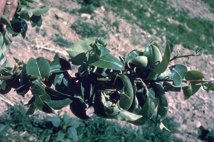
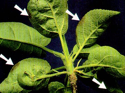
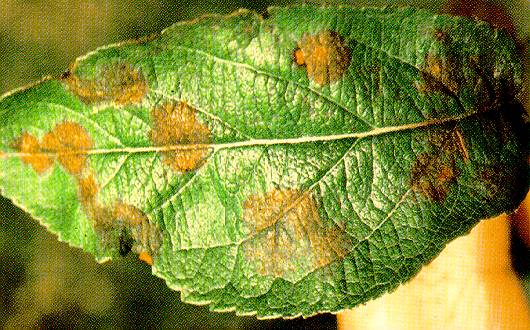
Alternaria_Blotch_1
Alternaria Blotch 1
Alternaria blotch on Delicious apple leaf.
(Alan L. Jones, Michigan State University and Turner B. Sutton, North Carolina State University)
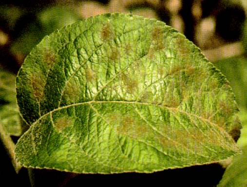
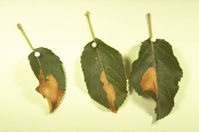
Alternaria_Blotch_2
Alternaria Blotch 2
Progressive development of Alternaria blotch across leaves on a shoot of Delicious apple.
(Alan L. Jones, Michigan State University and Turner B. Sutton, North Carolina State University)
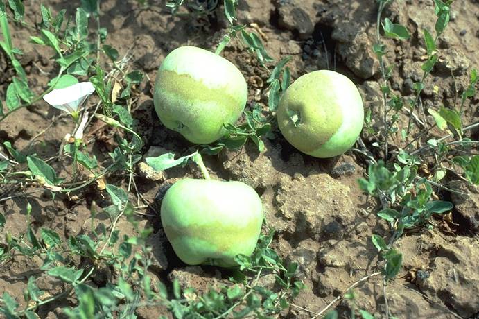
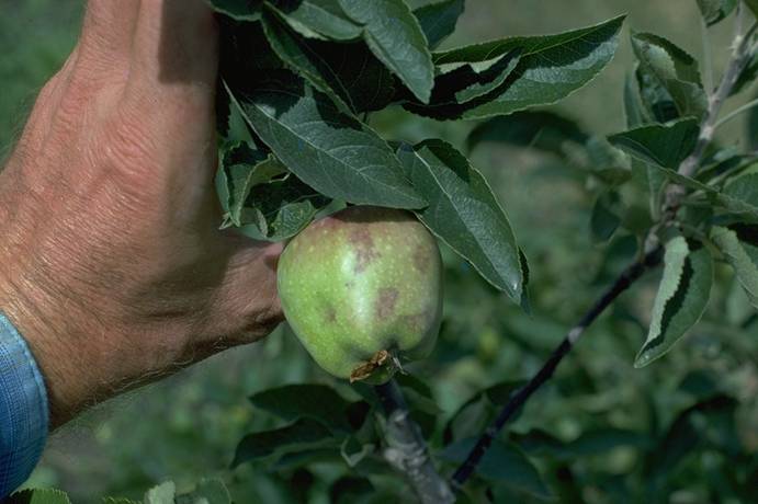
Apple_bitter_pit,_calcium_deficiency
Apple bitter pit, calcium deficiency (H. J. Larsen; Colorado State University)
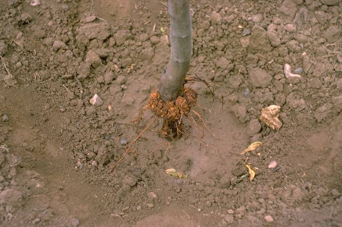
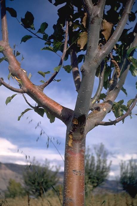
Apple_crown_gall
Apple crown gall (N. S. Luepschen; Colorado State University)
Apple_crown_gall
Apple crown gall (N. S. Luepschen; Colorado State University)
Apple_maggot_damage,_external_appearance_of_fruit
Apple maggot damage, external appearance of fruit (H. J. Larsen; Colorado State University)
Apple_maggot_damage,_internal_&_external_appearance_of_fruit
Apple maggot damage, internal & external appearance of fruit (H. J. Larsen; Colorado State University)
Apple_maggot_fruit_damage
Apple maggot fruit damage (H. J. Larsen; Colorado State University)
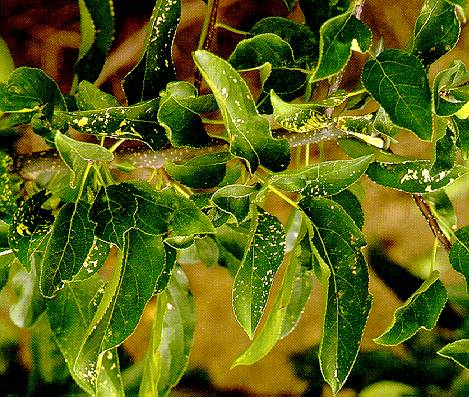
Apple_Mosaic
Apple Mosaic
Creamy-yellow areas in apple leaves infected with apple mosaic virus.
(Alan L. Jones, Michigan State University and Turner B. Sutton, North Carolina State University)
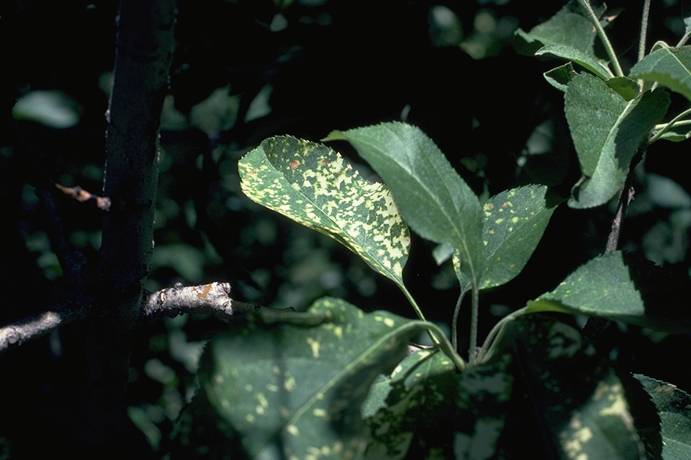
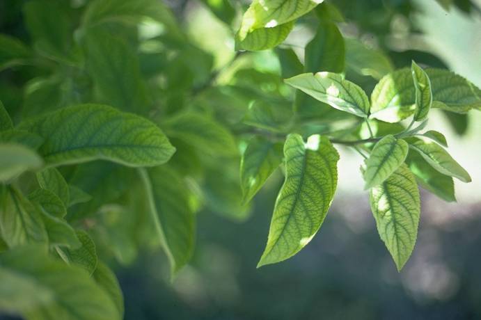
Apple_mosaic_virus,_leaf_symptoms_on_Golden_Delicious
Apple mosaic virus, leaf symptoms on Golden Delicious (H. J. Larsen; Colorado State University)
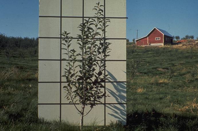
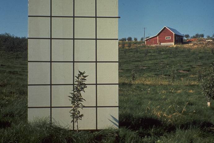
Apple_replant_disease_(ARD),_response_to_fall_preplant_fumigation
Apple replant disease (ARD), response to fall preplant fumigation (R. P. Covey; Colorado State University)
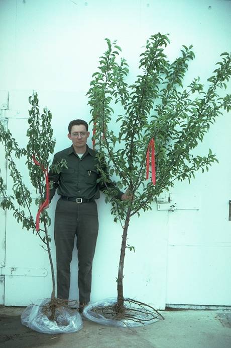
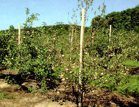
Apple_replant_problem,_left_non-fumigated,_right_fumigated
Apple replant problem, left non-fumigated, right fumigated (P. Burts; Colorado State University)
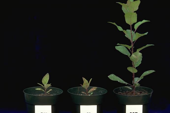
Apple_replant_study_-_ARD_bioassay_results_@_76_days
Apple replant study - ARD bioassay results @ 76 days (H. J. Larsen; Colorado State University)
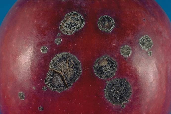
Apple_scab_fruit_infection
Apple scab fruit infection (P. Burts; Colorado State University)
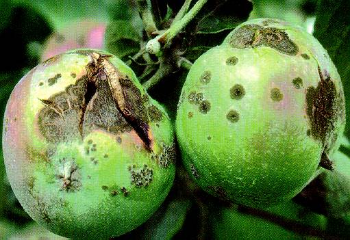
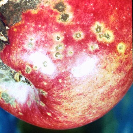
Apple_scab_lesion_1
Apple scab lesion 1
Apple scab lesions on fruit and leaves near midseason.
(Alan L. Jones, Michigan State University and Turner B. Sutton, North Carolina State University)
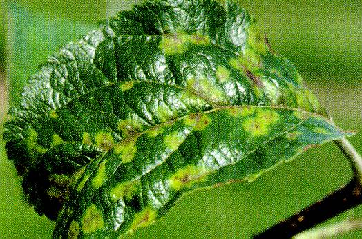
Apple_scab_lesion_2
Apple scab lesion 2
Primary apple scab lesions on the undersides of McIntosh apple leaves.
(Alan L. Jones, Michigan State University and Turner B. Sutton, North Carolina State University)
Apple_scab_lesion_3
Apple scab lesion 3
Sporulation of Venturia inaequalis in scab lesions on a McIntosh apple leaf.
(Alan L. Jones, Michigan State University and Turner B. Sutton, North Carolina State University)
Apple_scab_lesion_4
Apple scab lesion 4
Primary scab infection on the blossom end (calyx) of a McIntosh apple.
(Alan L. Jones, Michigan State University and Turner B. Sutton, North Carolina State University)
Apple_scab_lesion_5
Apple scab lesion 5
Cracking of McIntosh fruit associated with large scab lesions late in the season.
(Alan L. Jones, Michigan State University and Turner B. Sutton, North Carolina State University)
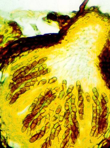
Apple_scab_lesion_6
Apple scab lesion 6
Cross-section of a pseudothecium of the apple scab fungus with pigmented mature ascospores in rows of eight ascospores per ascus.
(Alan L. Jones, Michigan State University and Turner B. Sutton, North Carolina State University)
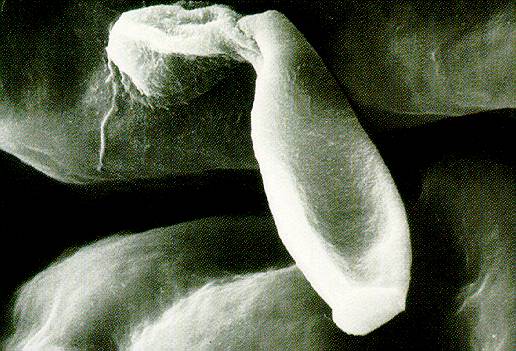
Apple_scab_lesion_7
Apple scab lesion 7
Germination of a conidium of the apple scab fungus. The spore sends out a short germ tube with an appressorium at its tip from which the fungus penetrates the host tissue. Ascospores germinate similarly.
(Alan L. Jones, Michigan State University and Turner B. Sutton, North Carolina State University)
Apple_scab_lesion_8
Apple scab lesion 8
Presymptom scab control. Chlorotic apple scab lesions produced when a sterol demethylation inhibitor was applied after infection but before symptoms were observed.
(Alan L. Jones, Michigan State University and Turner B. Sutton, North Carolina State University)
Apple_scab_lesion_9
Apple scab lesion 9
Post symptom scab control. Necrotic apple scab lesions produced when a dodine-captan combination was applied to control visible infections.
(Alan L. Jones, Michigan State University and Turner B. Sutton, North Carolina State University)
Apple_Union_Necrosis_and_Decline_1
Apple Union Necrosis and Decline 1
Typical weak growth and pale coloration of foliage of Delicious on Mm.106 rootstock with apple union necrosis and decline.
(Alan L. Jones, Michigan State University and Turner B. Sutton, North Carolina State University)
Apple_Union_Necrosis_and_Decline_2
Apple Union Necrosis and Decline 2
Line of dead tissue at the union between the Delicious scion and the MM.106 rootstock is typical of union necrosis and decline.
(Alan L. Jones, Michigan State University and Turner B. Sutton, North Carolina State University)
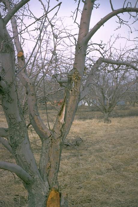
Apple_Union_Necrosis_and_Decline_3
Apple Union Necrosis and Decline 3
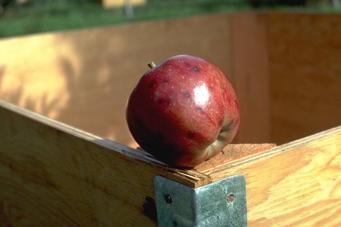
Breaking off of apple tree at the union between rootstock and scion is typical of union necrosis and decline.
(Alan L. Jones, Michigan State University and Turner B. Sutton, North Carolina State University)
Apple_winter_injury
Apple winter injury (N. S. Luepschen; Colorado State University)
ARD_symptoms_of_poor_growth_on_2nd_leaf_tree,_non-fumigated
ARD symptoms of poor growth on 2nd leaf tree, non-fumigated (R. P. Covey; Colorado State University)
Bitter_Rot_
Bitter Rot
Bitter_Rot_1
Bitter Rot 1
Golden Delicious apple with bitter rot lesion. Note the sunken nature and presence of acervuli with salmon or pink spores.
(Alan L. Jones, Michigan State University and Turner B. Sutton, North Carolina State University)
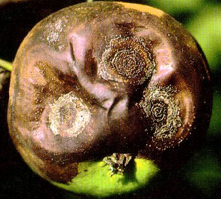
Bitter_Rot_2
Bitter Rot 2
Later stage of bitter rot. Note depressed lesions with concentric rings of acervuli and conidia.
(Alan L. Jones, Michigan State University and Turner B. Sutton, North Carolina State University)
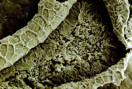
Bitter_Rot_3
Bitter Rot 3
Conidia of Colletotrichum gloeosporioides within an acervulus.
(Alan L. Jones, Michigan State University and Turner B. Sutton, North Carolina State University)
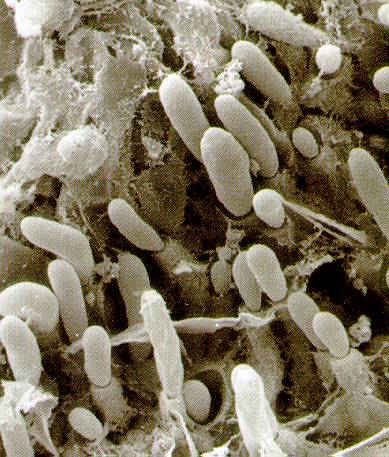
Bitter_Rot_4
Bitter Rot 4
Conidia of Colletotrichum gloeosporioides extruding from phialides within the acervulus.
(Alan L. Jones, Michigan State University and Turner B. Sutton, North Carolina State University)
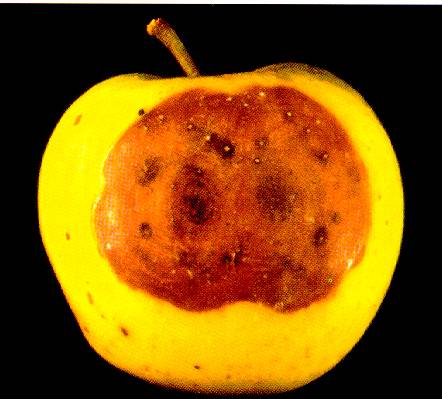
Bitter_Rot_5
Bitter Rot 5
Golden Delicious apple infected with a perithecial strain of the bitter rot fungus. Lesions are usually not sunken and are often darker than those caused by conidial strains.
(Alan L. Jones, Michigan State University and Turner B. Sutton, North Carolina State University)
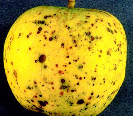
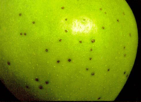
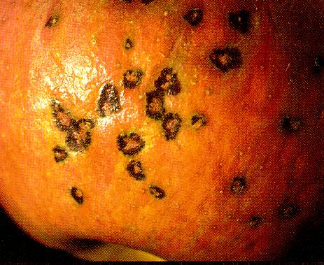
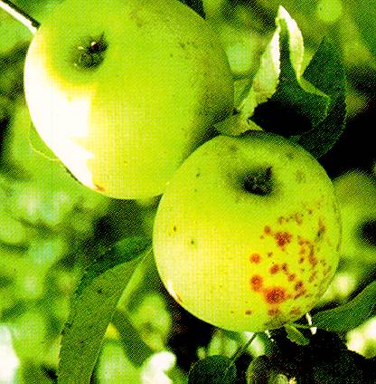
Black_Pox
Black Pox
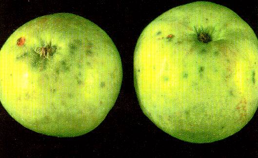
Small black spots of black pox, caused by Helminthosporium papulosum, on Grimes Golden apples.
(Alan L. Jones, Michigan State University and Turner B. Sutton, North Carolina State University)
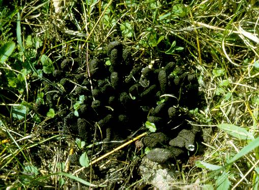
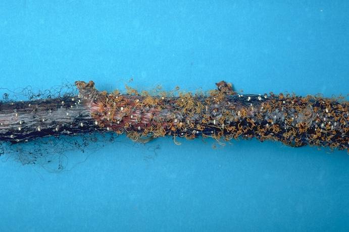
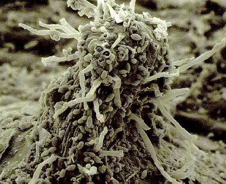
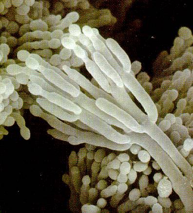
Black_Root_Rot
Black Root Rot
Finger-like fruiting structures of the black root rot pathogen Xylaria mali.
(Alan L. Jones, Michigan State University and Turner B. Sutton, North Carolina State University)
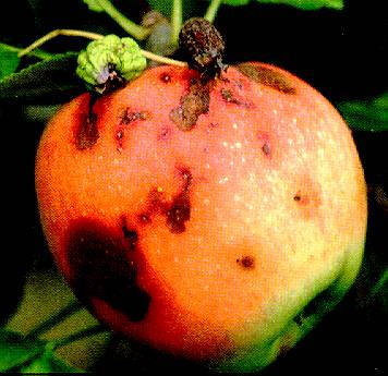
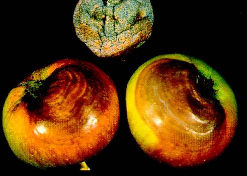
Black_Rot_
Black Rot
Black_Rot_1
Black Rot 1
Initial stages of black rot. Note necrotic spots developing below mummified fruit.
(Alan L. Jones, Michigan State University and Turner B. Sutton, North Carolina State University)
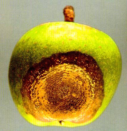
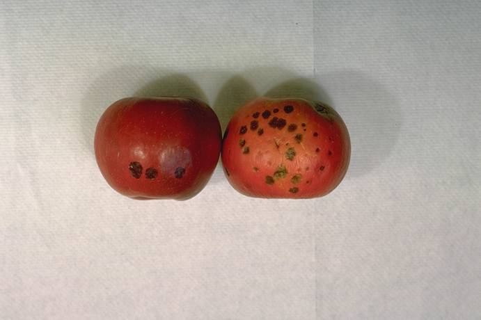
Black_Rot_2
Black Rot 2
Two Cortland apples with black rot. Note brown, wavy patterns developing at the calyx end and a mummified fruit with pycnidia.
(Alan L. Jones, Michigan State University and Turner B. Sutton, North Carolina State University)
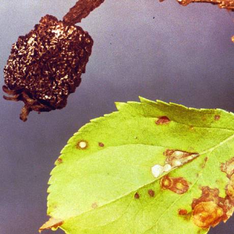
Black_Rot_Mummy_and_Frog-Eye_Leaf_Spot_
Black Rot Mummy and Frog-Eye Leaf Spot
Blister_bark_3
Blister bark on delicious apple (C. L. Parish, USDA)
Blister_Spot_1
Blister Spot 1
Shiny, raised blister spot lesions on Mutsu apple.
(Alan L. Jones, Michigan State University and Turner B. Sutton, North Carolina State University)
Blister_Spot_2
Blister Spot 2
Expanded blister spot lesions on mature Cortland apple.
(Alan L. Jones, Michigan State University and Turner B. Sutton, North Carolina State University)
Blister_Spot_3
Blister Spot 3
Shoot of Mutsu with ooze droplet containing blister spot bacteria and curled leaves due to infection on the midvein.
(Alan L. Jones, Michigan State University and Turner B. Sutton, North Carolina State University)
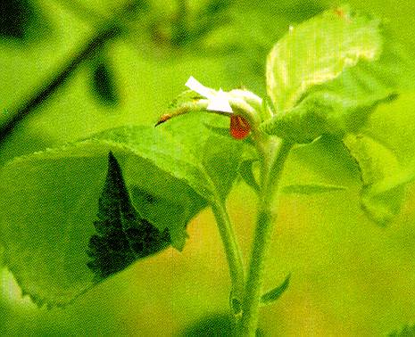
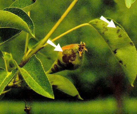
Blossom_Blast_of_Pear
Blossom Blast of Pear
Blossom blast of pear. Note death of spur but failure of shoot to become infected.
(Alan L. Jones, Michigan State University and Turner B. Sutton, North Carolina State University)
Blotch
Blotch
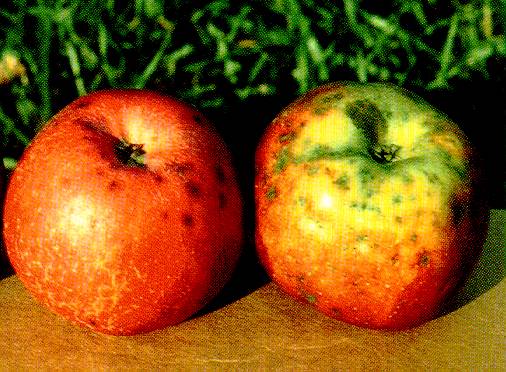
Apple blotch on the fruit.
(Alan L. Jones, Michigan State University and Turner B. Sutton, North Carolina State University)
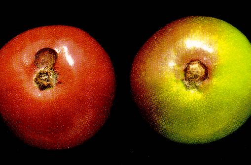
Brooks_Spot_1
Brooks Spot 1
Brooks spot on Jonathan apple fruit.
(Alan L. Jones, Michigan State University and Turner B. Sutton, North Carolina State University)
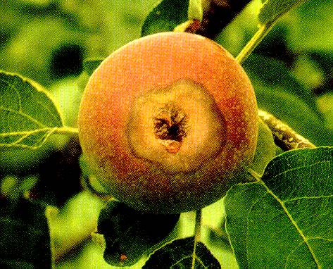
Brooks_Spot_2
Brooks Spot 2
Brooks spot on the calyx end of Grimes Golden apples.
(Alan L. Jones, Michigan State University and Turner B. Sutton, North Carolina State University)
Brooks_Spot_3
Brooks Spot 3
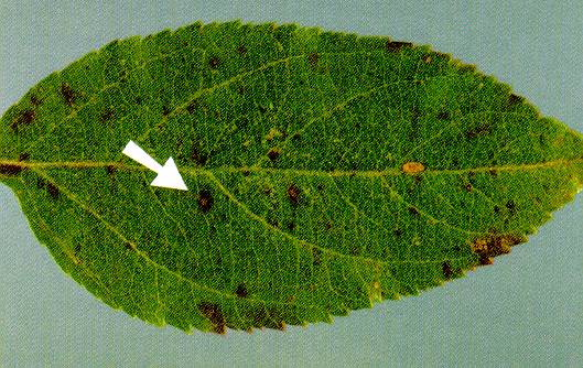
Small purple spots on an apple leaf caused by the Brooks spot fungus.
(Alan L. Jones, Michigan State University and Turner B. Sutton, North Carolina State University)
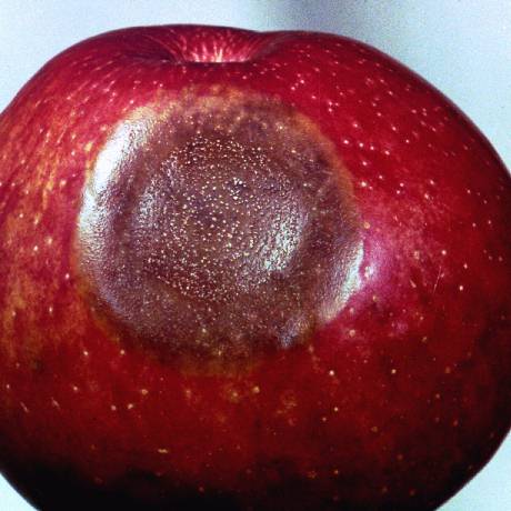
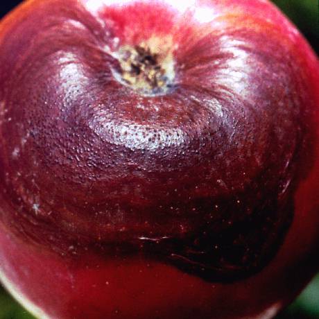
Bull's-eye_Rot
Bull's-eye Rot
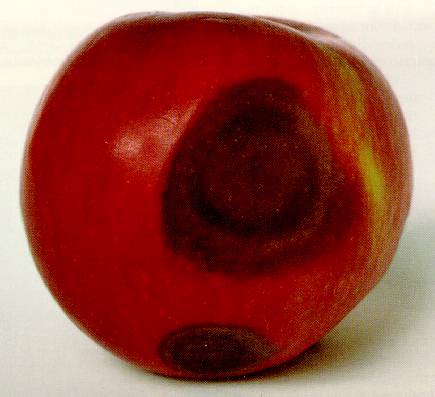
Bull's-eye rot on McIntosh apple. Note light color in center of depressed lesion and alternating areas of tan and brown.
(Alan L. Jones, Michigan State University and Turner B. Sutton, North Carolina State University)
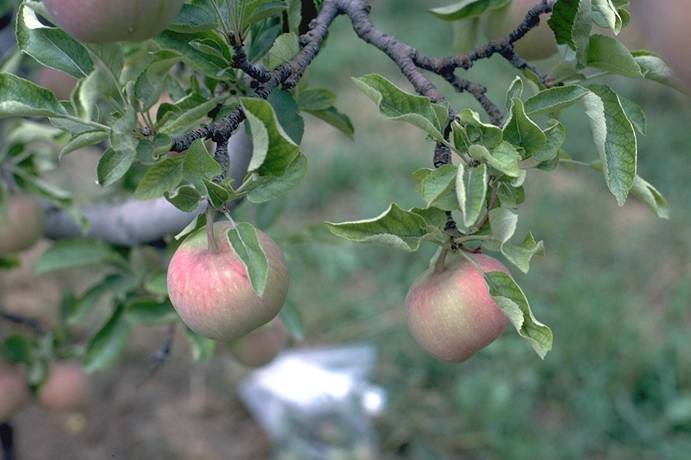
Calcium_deficiency,_Jonathan_spot
Calcium deficiency, Jonathan spot (N. S. Luepschen; Colorado State University)
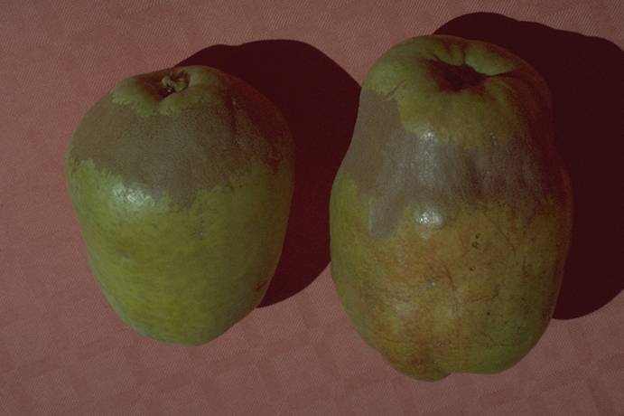
Calcium_deficiency_corky_spot_on_d'_Anjou_pear
Calcium deficiency corky spot on d' Anjou pear (N. S. Luepschen; Colorado State University)
Cedar_Apple_Rust_Gall
Cedar Apple Rust Gall
Cedar_Apple_Rust_on_Leaf_
Cedar Apple Rust on Leaf
Cherry_raspleaf_virus,_normal_vs_symptomatic_fruit
Cherry raspleaf virus, normal vs symptomatic fruit (C. L. Parish; Colorado State University)

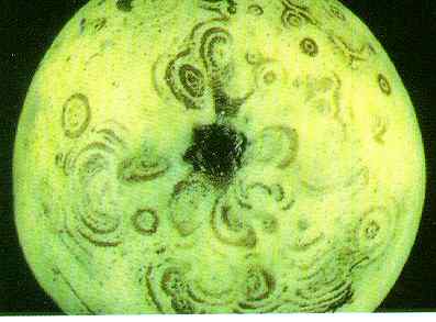
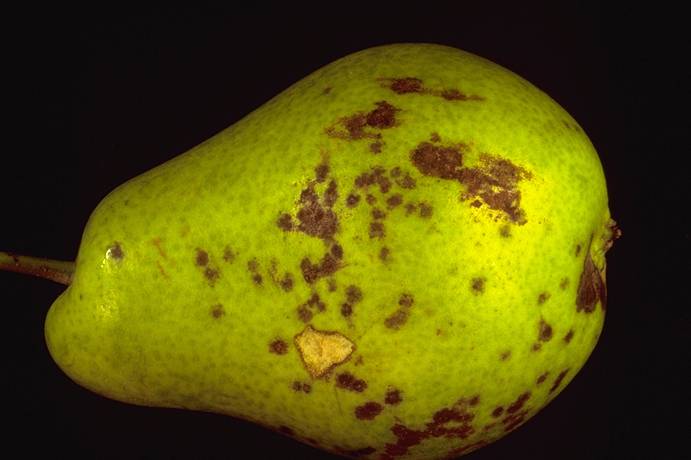
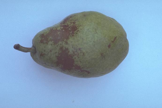
Concentric_ring_pattern_1
Concentric ring pattern on pear, calyx end (T. Van der Zwet, USDA)
Cycle_apple_scab
Cycle apple scab
Disease cycle of apple scab.
(Alan L. Jones, Michigan State University and Turner B. Sutton, North Carolina State University)

Cycle_blight
Cycle blight
Disease cycle of blight.
(Alan L. Jones, Michigan State University and Turner B. Sutton, North Carolina State University)

Cycle_powdery_mildew
Cycle powdery mildew
Disease cycle of powdery mildew.
(Alan L. Jones, Michigan State University and Turner B. Sutton, North Carolina State University)

Cycle_rust
Cycle rust
Disease cycle of cedar-apple rust.
(Alan L. Jones, Michigan State University and Turner B. Sutton, North Carolina State University)

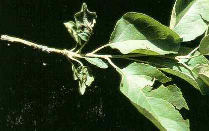
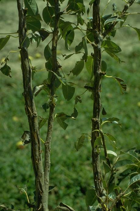
Cytospora_canker_dieback_on appple
Cytospora canker dieback on apple (H. J. Larsen; Colorado State University)
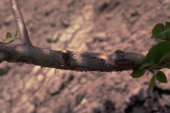
Cytospora_canker_infection_growth_on_apple
Cytospora canker infection growth on apple (H. J. Larsen; Colorado State University)
Cytospora_canker_lesions_on_apple
Cytospora canker lesions on apple (H. J. Larsen; Colorado State University)
Cytospora_canker_of_apple
Cytospora canker of apple (N. S. Luepschen; Colorado State University)
Cytospora_spore_threads_on_apple
Cytospora spore threads on apple (P. Burts; Colorado State University)
Dry-Eye_and_Calyx-end_Rots_1
Dry-Eye and Calyx-end Rots 1
Dry-eye rot, caused by Botrytis cinerea, on McIntosh apple.
(Alan L. Jones, Michigan State University and Turner B. Sutton, North Carolina State University)
Dry-Eye_and_Calyx-end_Rots_2
Dry-Eye and Calyx-end Rots 2
Calyx-end rot on Paulared apple.
(Alan L. Jones, Michigan State University and Turner B. Sutton, North Carolina State University)
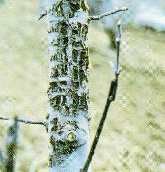
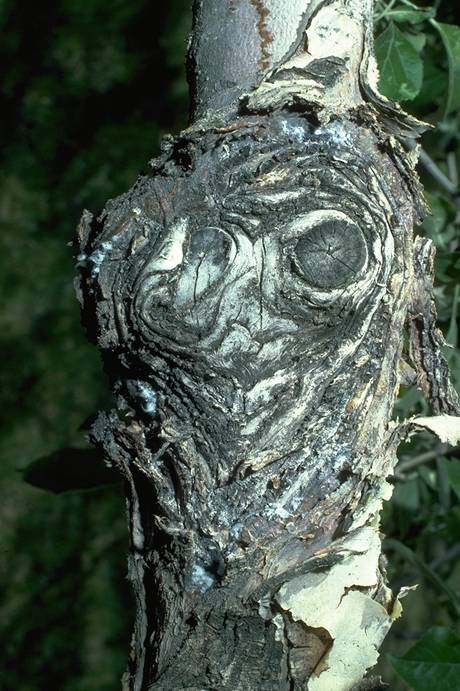
European_canker_(Nectria_galligena)_fruiting_bodies_on_apples
European canker (Nectria galligena) fruiting bodies on apples (H. J. Larsen; Colorado State University)
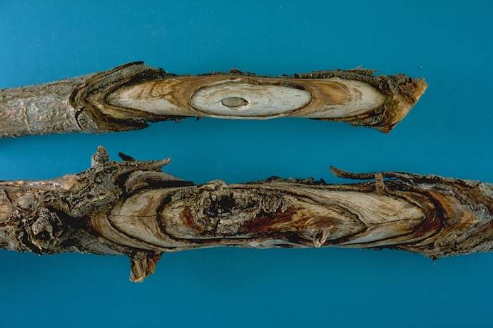
European_canker_on_apple
European canker on apple (H. J. Larsen; Colorado State University)
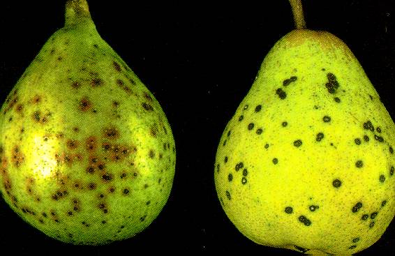
Fabraea_Leaf_Spot_1
Fabraea Leaf Spot 1
Fruit of Tennessee Selection #345298 (left) and Bartlett pear (right) infected by the Fabraea leaf spot pathogen.
(Alan L. Jones, Michigan State University and Turner B. Sutton, North Carolina State University)
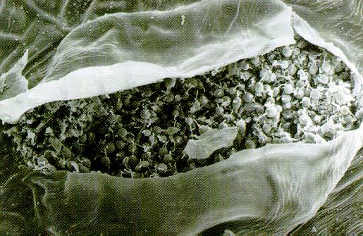
Fabraea_Leaf_Spot_2
Fabraea Leaf Spot 2
Surface view of an acervulus of Entomosporium maculatum breaking through the epidermis.
(Alan L. Jones, Michigan State University and Turner B. Sutton, North Carolina State University)
Fabraea_Leaf_Spot_3
Fabraea Leaf Spot 3
A conidium of Fabraea leaf spot with two lateral setae or hair-like structures attached to the cells.
(Alan L. Jones, Michigan State University and Turner B. Sutton, North Carolina State University)
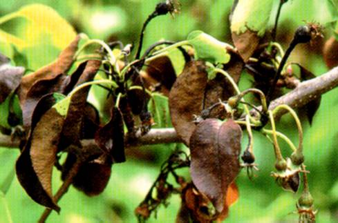
Fire_Blight
Fire Blight
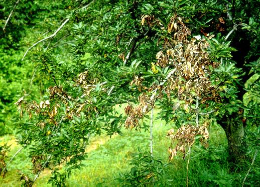
Fire_blight_1
Fire blight 1
Apple tree with dieback of spurs and shoots caused by fire blight.
(Alan L. Jones, Michigan State University and Turner B. Sutton, North Carolina State University)
Fire_blight_2
Fire blight 2
First symptoms of blossom blight. Note ooze droplets and discoloration of the blossom on the lower right.
(Alan L. Jones, Michigan State University and Turner B. Sutton, North Carolina State University)
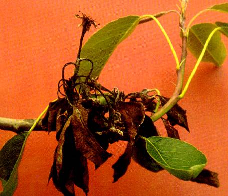
Fire_blight_3
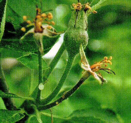
Fire blight 3
Fruit spurs on pear with fire blight-infected blossoms.
(Alan L. Jones, Michigan State University and Turner B. Sutton, North Carolina State University)
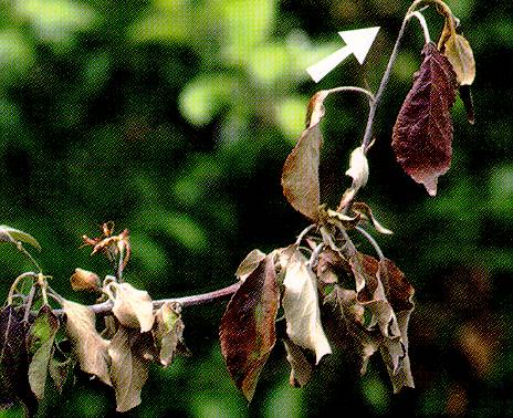
Fire_blight_4
Fire blight 4
Apple shoot with fire blight. Note crook at tip.
(Alan L. Jones, Michigan State University and Turner B. Sutton, North Carolina State University)
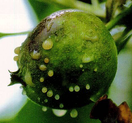
Fire_blight_5
Fire blight 5
Bacterial ooze exuding from an apple infected with fire blight.
(Alan L. Jones, Michigan State University and Turner B. Sutton, North Carolina State University)
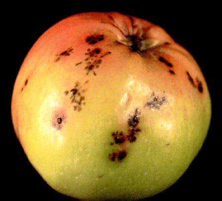
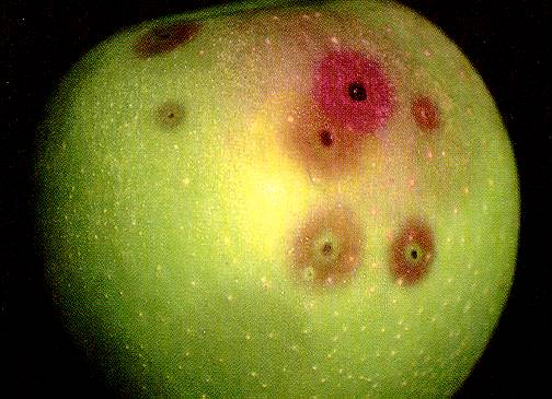
Fire_blight_6
Fire blight 6
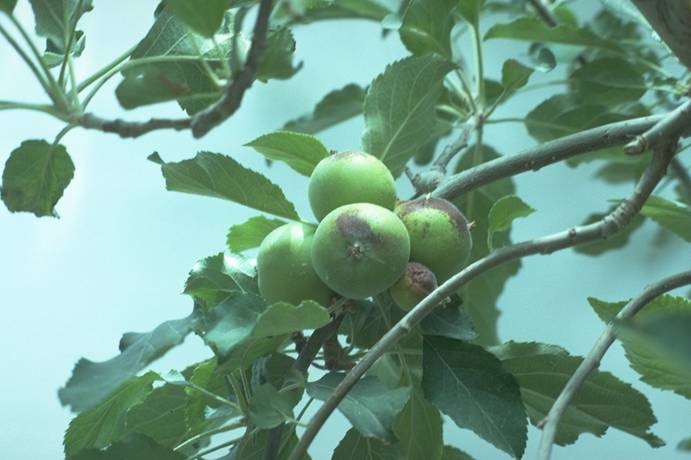
Necrotic spots with red margins on apple caused by fire blight. Infection associated with a severe storm about 8 weeks after bloom.
(Alan L. Jones, Michigan State University and Turner B. Sutton, North Carolina State University)
Fire_blight_7
Fire blight 7
Decline of Spy apple trees due to infection of the Mark rootstock by fire blight.
(Alan L. Jones, Michigan State University and Turner B. Sutton, North Carolina State University)
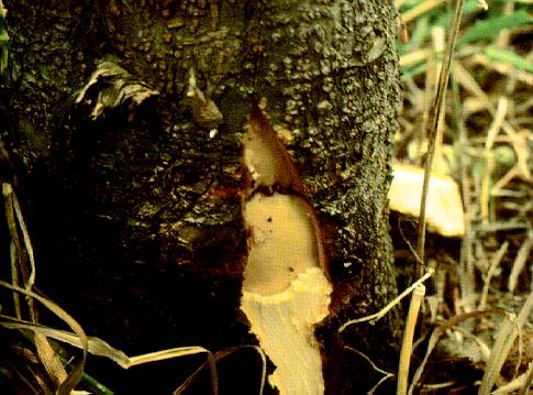
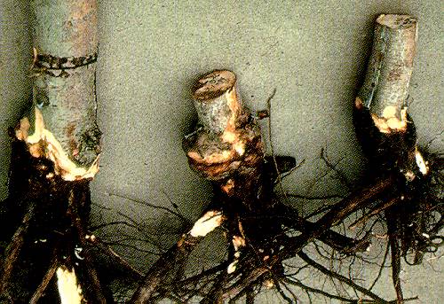
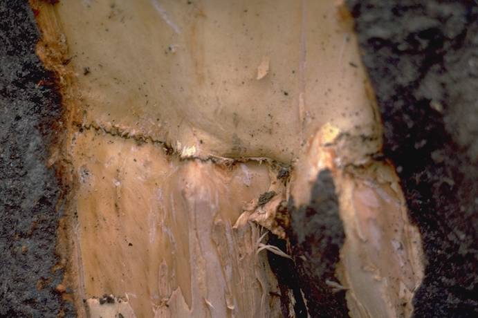
Fire_blight_8
Fire blight 8
Fire blight on apple rootstocks just below ground level. Bark cut away to show the margin between healthy and diseased crown and root tissues.
(Alan L. Jones, Michigan State University and Turner B. Sutton, North Carolina State University)
Fire_blight_canker_on_pear
Fire blight canker on pear (N. S. Luepschen; Colorado State University)
Fire_blight_canker_with_ooze_on_pear
Fire blight canker with ooze on pear (N. S. Luepschen; Colorado State University)
Fire_blight_infection_&_scaffold_loss_of_apple
Fire blight infection & scaffold loss of apple (C. E. Swift; Colorado State University)
Fire_blight_on_2bartlett_pear_
Fire blight on bartlett pear (N. S. Luepschen; Colorado State University)
Fire_blight_on_pear_shoots
Fire blight on pear shoots (N. S. Luepschen; Colorado State University)
Flat_apple_(cherry_raspleaf_virus)_tree_symptoms_(growth_pattern)
Flat apple (cherry raspleaf virus) tree symptoms (growth pattern)
(H. J. Larsen; Colorado State University)
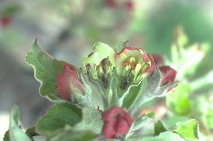
Freeze_damage_to_apple_flowers
Freeze damage to apple flowers (A. R. Renquist; Colorado State University)
Freeze_damage_to_apple_flowers
Freeze damage to apple flowers (A. R. Renquist; Colorado State University)
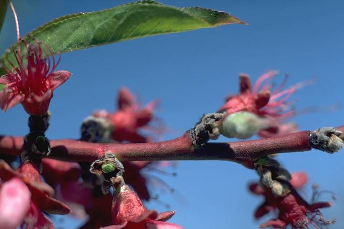
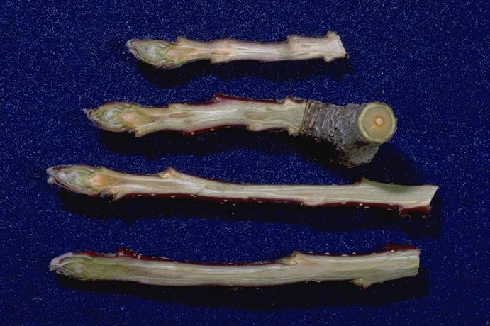
Freeze_damage_to_bartlett_pear_shoots
Freeze damage to bartlett pear shoots (H. J. Larsen; Colorado State University)
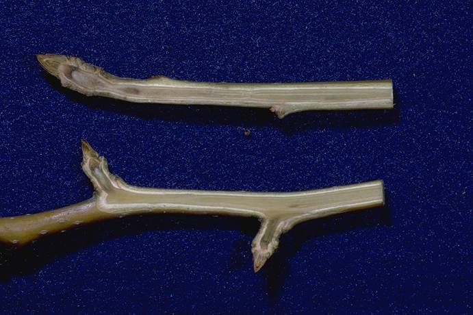
Freeze_injury_to_apple_shoots
Freeze injury to apple shoots (H. J. Larsen; Colorado State University)
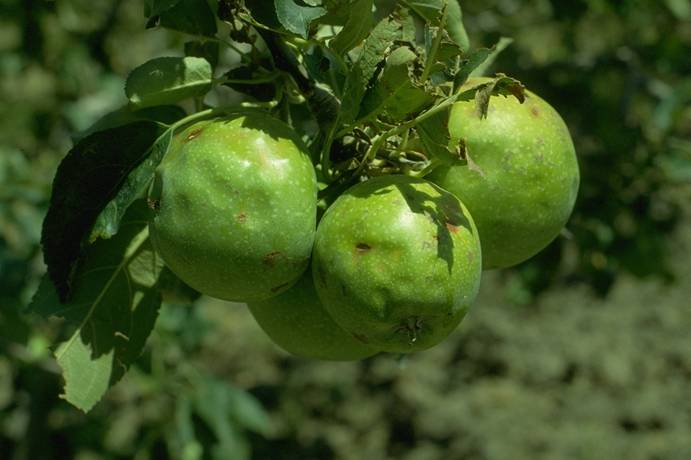
Frost_injury/hail_injury
Spring frost injury and hail injury S. V. Thomson, Utah State University
Golden_apple,_northwestern_anthracnose_canker
Golden apple, northwestern anthracnose canker (H. J. Larsen; Colorado State University)
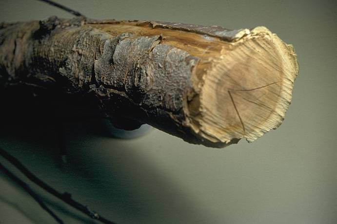
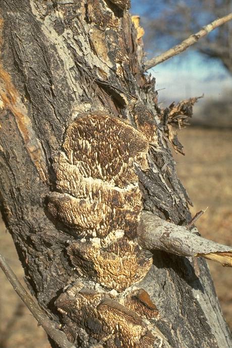
Golden_Delicious_apple,_apple_wood_rot,_(Trametes_hispida)
Golden Delicious apple, apple wood rot, (Trametes hispida)
(N. S. Luepschen; Colorado State University)
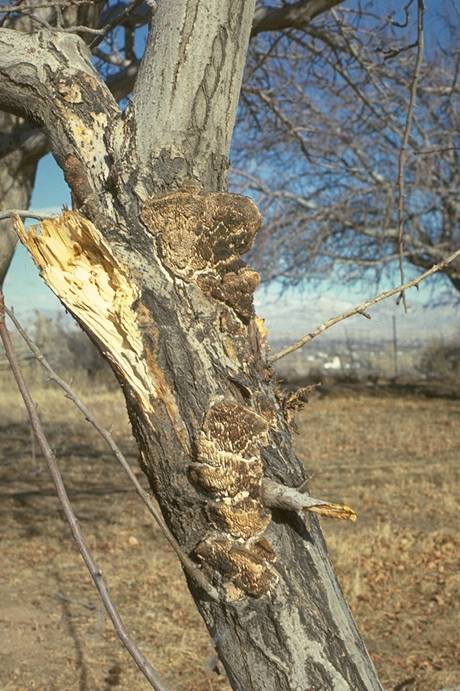
Golden_Delicious_apple,_apple_wood_rot_(Trametes_hispida)
Golden Delicious apple, apple wood rot (Trametes hispida)
(N. S. Luepschen; Colorado State University)
Golden_Delicious_apple_frost_injury
Golden Delicious apple frost injury (A. R. Renquist; Colorado State University)
Golden_Delicious_apple_iron_chlorosis_(deficiency)_leaf_symptoms
Golden Delicious apple iron chlorosis (deficiency) leaf symptoms (A. R. Renquist; Colorado State University)
Gray_Mold
Gray Mold
A "nest" of apples with gray mold caused by Botrytis.
(Alan L. Jones, Michigan State University and Turner B. Sutton, North Carolina State University)
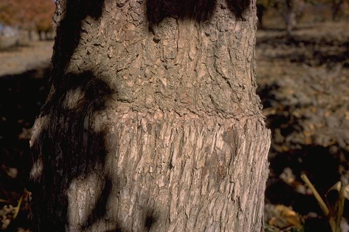
Hail_damage_on_pear_tree_bark
Hail damage on pear tree bark (H. J. Larsen; Colorado State University)
Hail_damage_to_apple_fruit
Hail damage to apple fruit (H. J. Larsen; Colorado State University)
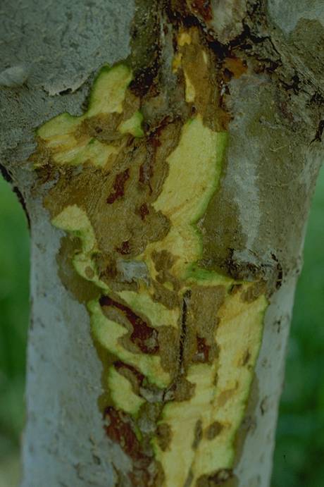
Jonathan_apple_slime_flux_kill,_cambium_injury_symptoms
Jonathan apple slime flux kill, cambium injury symptoms (N. S. Luepschen; Colorado State University)
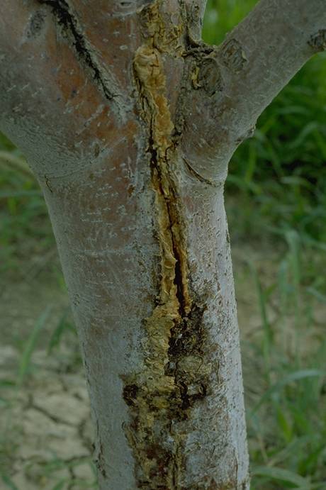
Jonathan_apple_slime_flux_on_lower_trunk
Jonathan apple slime flux on lower trunk (N. S. Luepschen; Colorado State University)
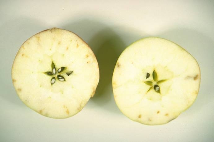
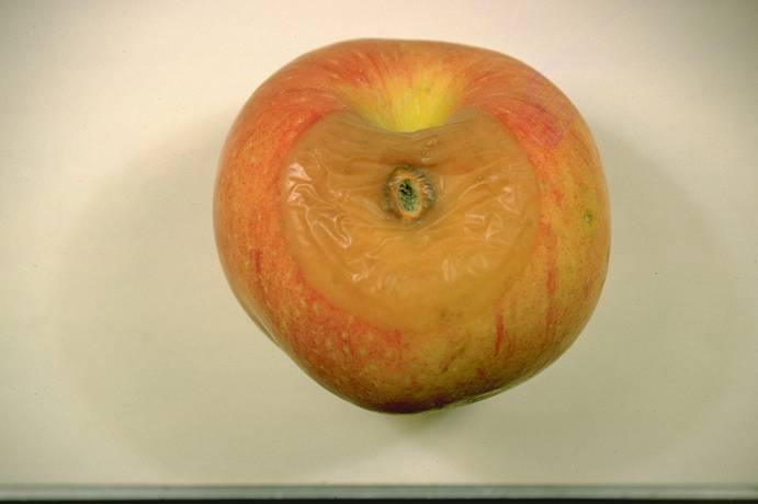
Jonathan_spot,_internal_calcium_deficiency
Jonathan spot, internal calcium deficiency (C. R. Ure; Colorado State University)
Leucostoma_Canker_1
Leucostoma Canker 1
A Leucostoma canker on Delicious apple with characteristic rough, scaly bark.
(Alan L. Jones, Michigan State University and Turner B. Sutton, North Carolina State University)
Leucostoma_Canker_2
Leucostoma Canker 2
Dieback of a central leader of Delicious apple caused by Leucostoma canker.
(Alan L. Jones, Michigan State University and Turner B. Sutton, North Carolina State University)
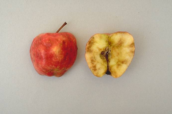
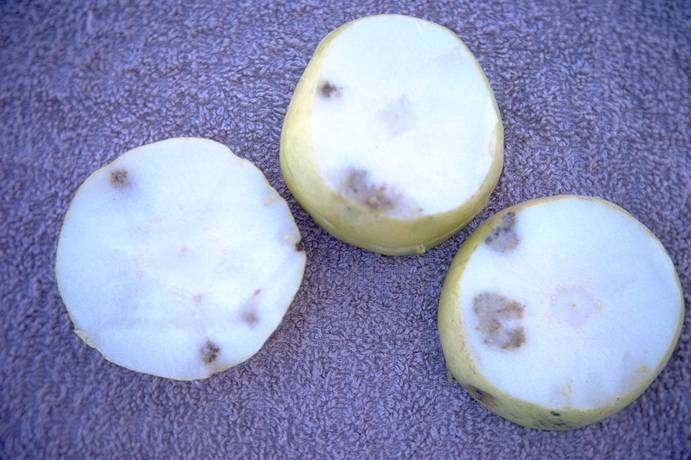
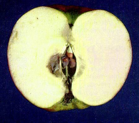
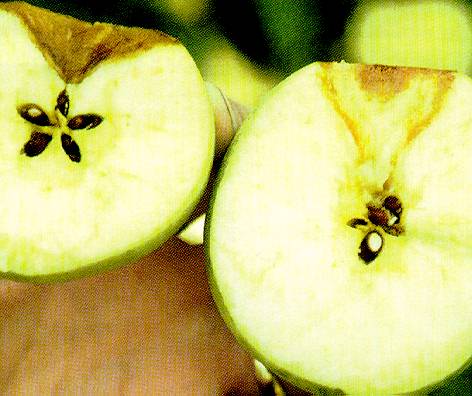
Moldy_Core_1
Moldy Core 1
Cross-section of apple with moldy core developing around the core region.
(Alan L. Jones, Michigan State University and Turner B. Sutton, North Carolina State University)
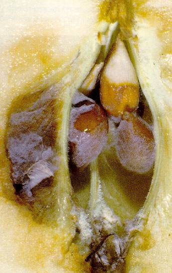
Moldy_Core_2
Moldy Core 2
Close-up of seed cavity overgrown with mold.
(Alan L. Jones, Michigan State University and Turner B. Sutton, North Carolina State University)
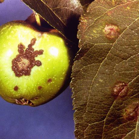
Mycosphaerella_Leaf_Spot
Mycosphaerella Leaf Spot
Mycosphaerella leaf spot on pear leaves and fruit.
(Alan L. Jones, Michigan State University and Turner B. Sutton, North Carolina State University)
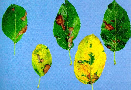
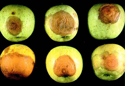
Necrotic_Leaf_Blotch_1
Necrotic Leaf Blotch 1
Golden Delicious leaves with necrotic leaf blotch. Early symptom development is shown in top row; greatly advanced stage in bottom row.
(Alan L. Jones, Michigan State University and Turner B. Sutton, North Carolina State University)
Necrotic_Leaf_Blotch_2
Necrotic Leaf Blotch 2
Leaf yellowing and defoliation of Golden Delicious from necrotic leaf blotch. The bottom leaves exhibit symptoms first, followed by upper leaves.
(Alan L. Jones, Michigan State University and Turner B. Sutton, North Carolina State University)
Nectria_Canker
Nectria Canker
Nectria canker on the trunk of Delicious apple.
(Alan L. Jones, Michigan State University and Turner B. Sutton, North Carolina State University)
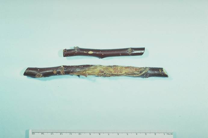
Nectria_Twig_Blight_1
Nectria Twig Blight 1
Dieback of Rome Beauty apple shoot from Nectria twig blight. Note canker at the base of the shoot.
(Alan L. Jones, Michigan State University and Turner B. Sutton, North Carolina State University)
Nectria_Twig_Blight_2a
Nectria Twig Blight 2a
Canker and orange fruiting structures of Nectira twig blight fungus.
(Alan L. Jones, Michigan State University and Turner B. Sutton, North Carolina State University)

Nectria_Twig_Blight_2b
Nectria Twig Blight 2b
Canker and orange fruiting structures of Nectira twig blight fungus.
(Alan L. Jones, Michigan State University and Turner B. Sutton, North Carolina State University)

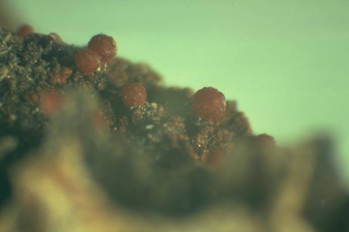
Nectria_Twig_Blight_3
Nectria Twig Blight 3
Orange fruiting structures of the Nectria twig blight fungus in a canker on McIntosh apple associated with a pruning wound.
(Alan L. Jones, Michigan State University and Turner B. Sutton, North Carolina State University)
Northwestern_anthracnose_canker_on_apple
Northwestern anthracnose canker on apple (H. J. Larsen; Colorado State University)
Orchard,_overhead_sprinkler_ice_damage
Orchard, overhead sprinkler ice damage (N. S. Luepschen; Colorado State University)
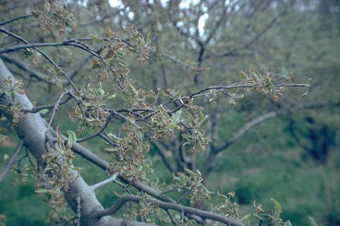
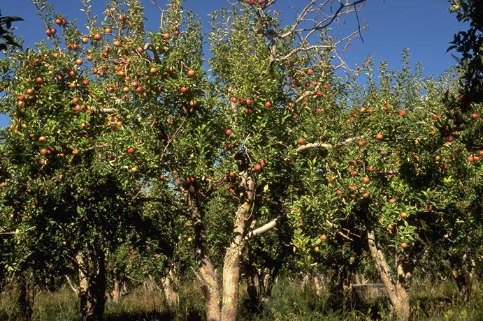
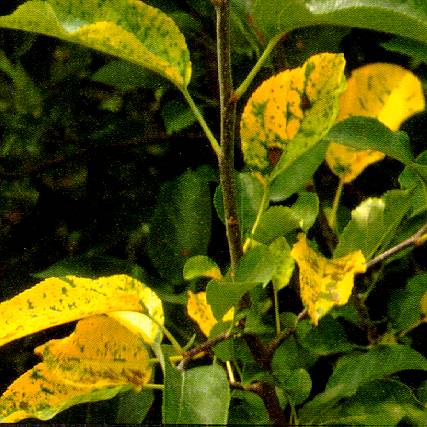
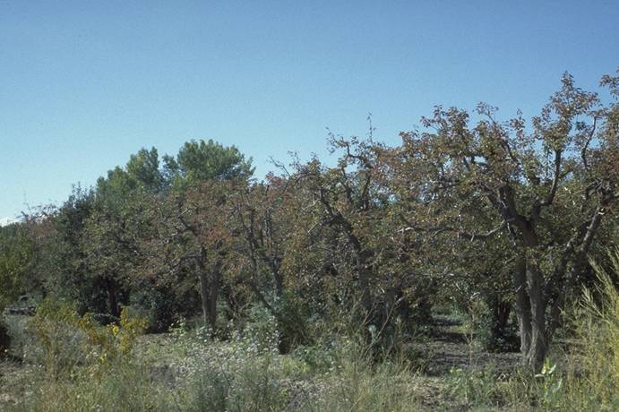
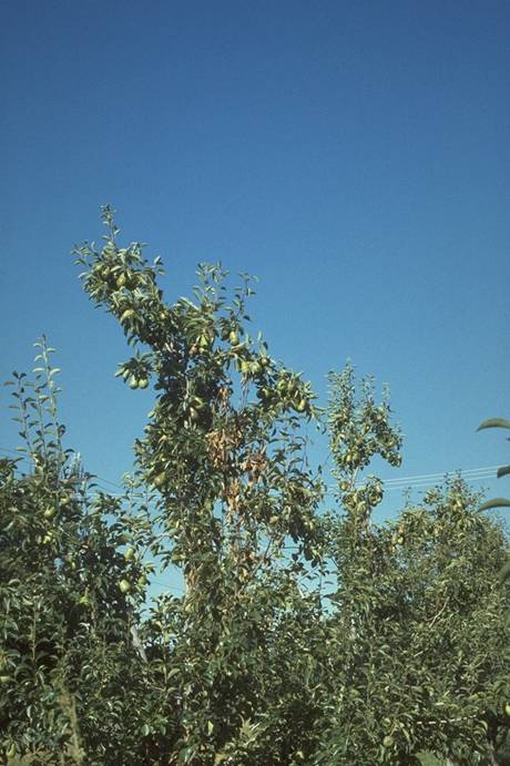
Pear_decline,_early_fall_leaf_coloration
Pear decline, early fall leaf coloration (H. J. Larsen; Colorado State University)
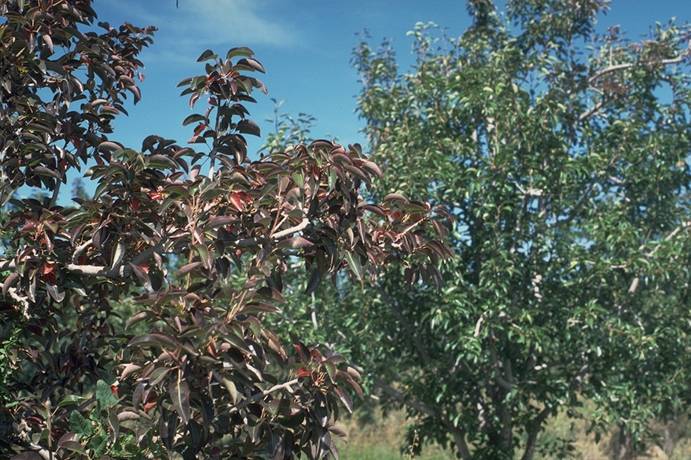
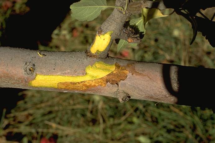
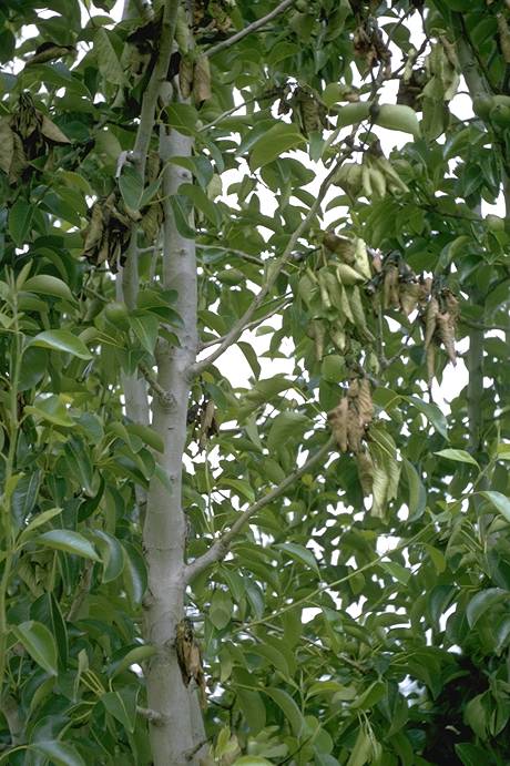
Pear_decline_(MLO)_branch_&_foliage_symptoms
Pear decline (MLO) branch & foliage symptoms (N. S. Luepschen; Colorado State University)
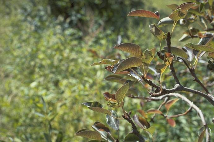
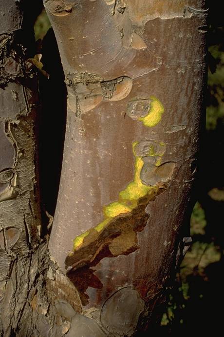
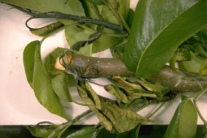
Pear_decline_(MLO)_branch_symptoms
Pear decline (MLO) branch symptoms (N. S. Luepschen; Colorado State University)
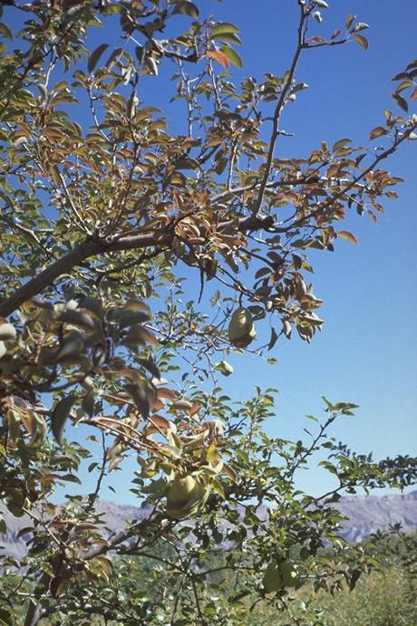
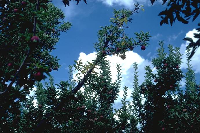
Pear_decline_(MLO)_growth_symptoms
Pear decline (MLO) growth symptoms (N. S. Luepschen; Colorado State University)
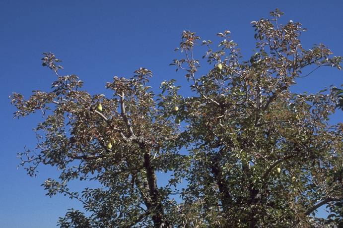
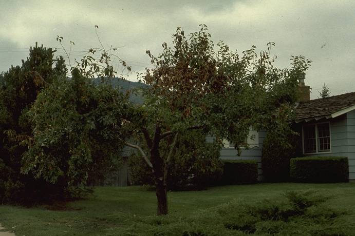
Pear_decline_(MLO)_whole_tree_symptoms
Pear decline (MLO) whole tree symptoms (N. S. Luepschen; Colorado State University)
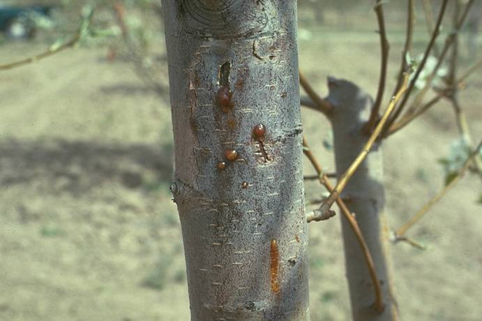
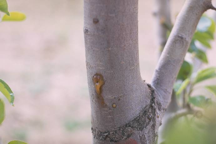
Pear_fire_blight_canker_with_ooze
Pear fire blight canker with ooze (N. S. Luepschen; Colorado State University)
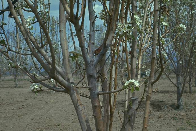
Pear_fire_blight_caused_by_hail_damage
Pear fire blight caused by hail damage (N. S. Luepschen; Colorado State University)
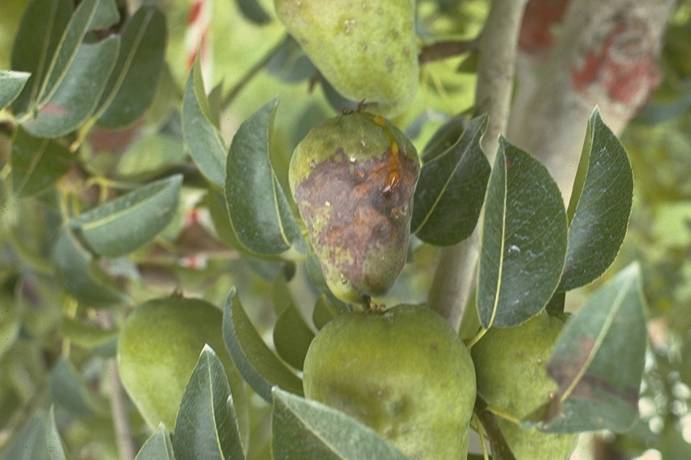
Pear_fire_blight_shoot_infection_+_ooze
Pear fire blight shoot infection + ooze (N. S. Luepschen; Colorado State University)
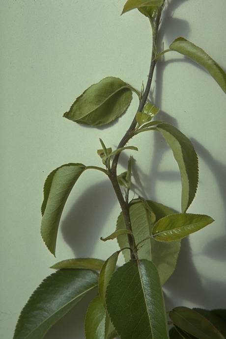
Pear_fire_blight_shoot_infection_symptoms
Pear fire blight shoot infection symptoms (N. S. Luepschen; Colorado State University)
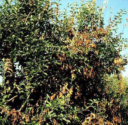
Pear_fire_blight_strikes_in_top_of_tree
Pear fire blight strikes in top of tree (N. S. Luepschen; Colorado State University)
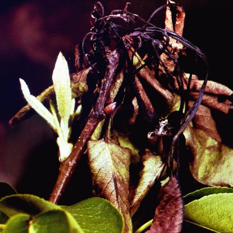
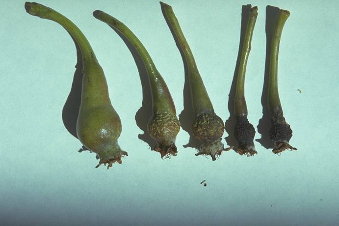
Pear_frost_damage
Pear frost damage (N. S. Luepschen; Colorado State University)
Pear_frost_injury
Pear frost injury (N. S. Luepschen; Colorado State University)
Pear_root_stock_union
Pear root stock union (N. S. Luepschen; Colorado State University)
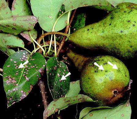
Pear_scab
Pear scab (L. E. Sommers; Colorado State University)
Pear_Scab_1
Pear Scab 1
Scab on young pear fruit and leaves.
(Alan L. Jones, Michigan State University and Turner B. Sutton, North Carolina State University)
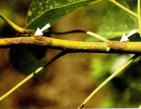
Pear_Scab_2
Pear Scab 2
Pear scab lesions on current-season shoot growth.
(Alan L. Jones, Michigan State University and Turner B. Sutton, North Carolina State University)
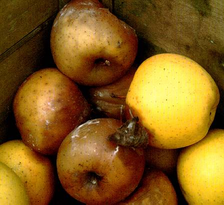
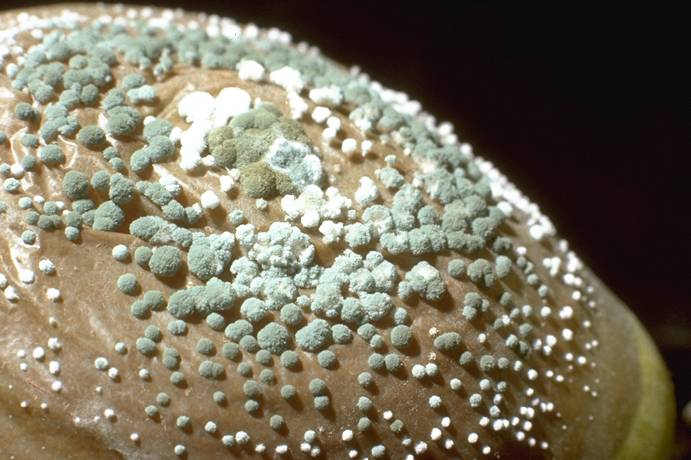
Penicillium_expansum_(blue_mold)_rot_on_pear_fruit
Penicillium expansum (blue mold) rot on pear fruit (H. J. Larsen; Colorado State University)
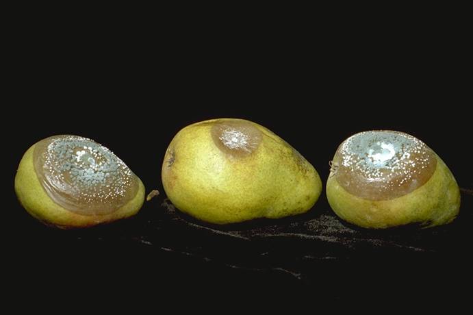
Penicillium_expansum_(blue_mold)_rot_on_pear_fruit
Penicillium expansum (blue mold) rot on pear fruit (H. J. Larsen; Colorado State University)
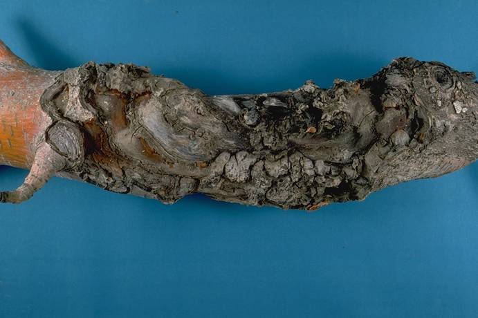
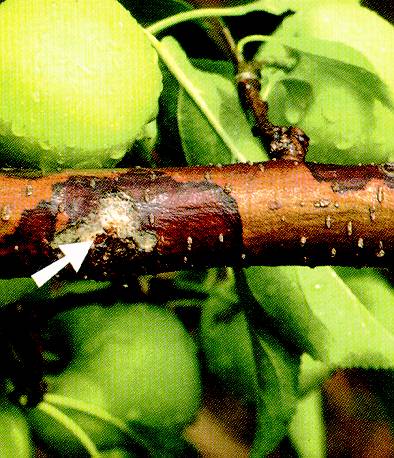
Perennial_canker_+_woolly_aphids_on_apple
Perennial canker + woolly aphids on apple (H. J. Larsen; Colorado State University)
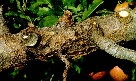
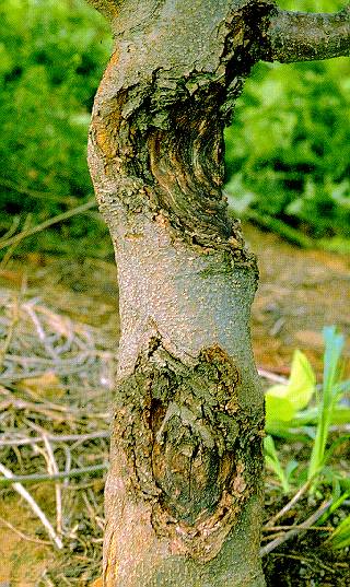
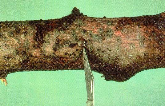
Perennial_canker_of_apple
Perennial canker of apple (H. J. Larsen; Colorado State University)
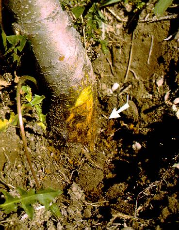
Phytophthora_collar_rot_symptoms
Phytophthora collar rot symptoms (N. S. Luepschen; Colorado State University)
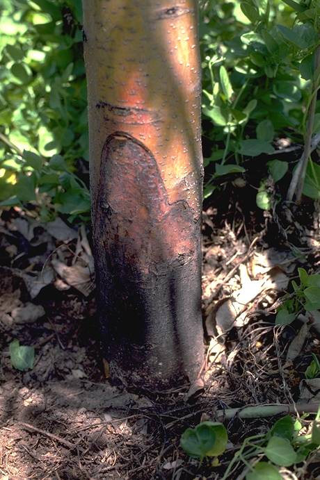
Phytophthora_crown_rot,_Early_Jonathan_trunk_canker
Phytophthora crown rot, Early Jonathan trunk canker (H. J. Larsen; Colorado State University)
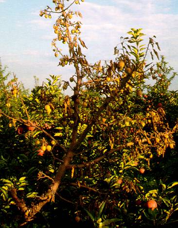
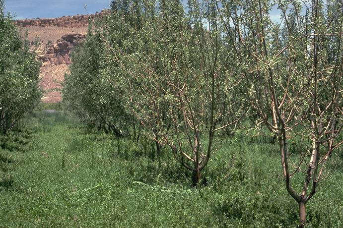
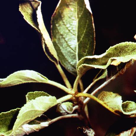
Phytophthora_crown_rot,_Erly_Jonathan_weak_tree_symptoms
Phytophthora crown rot, Erly Jonathan weak tree symptoms (H. J. Larsen; Colorado State University)
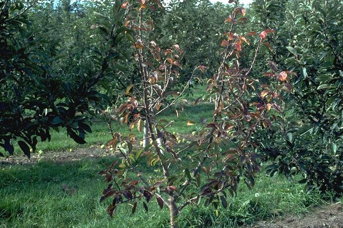
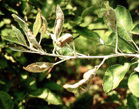
Phytophthora_crown_rot,_premature_fall_leaf_coloration
Phytophthora crown rot, premature fall leaf coloration (H. J. Larsen; Colorado State University)
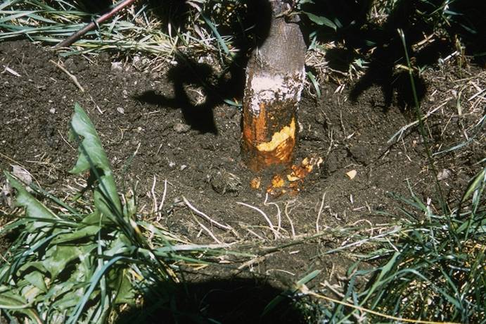
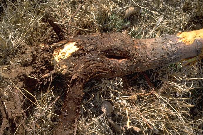
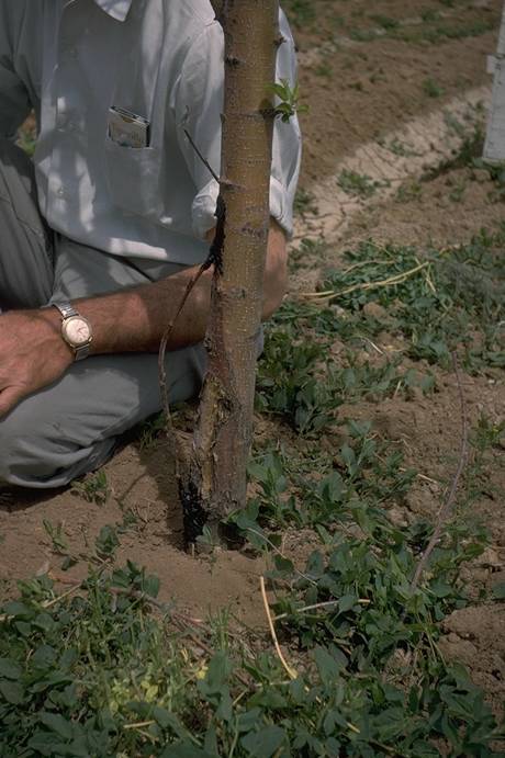
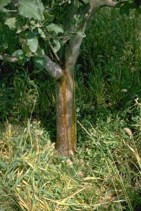
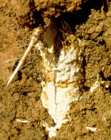
Phytophthora_Root,_Crown,_and_Collar_Rot
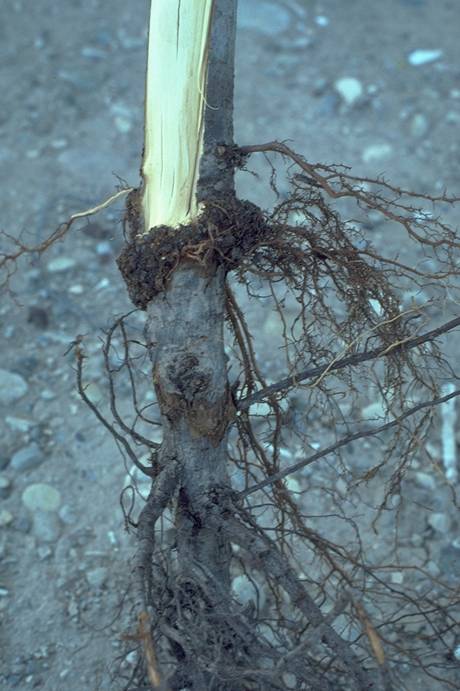
Phytophthora Root, Crown, and Collnar Rot
Brown decay of rootstock collar just below ground, a common symptom of Phytophthora root, crown and collar rot.
(Alan L. Jones, Michigan State University and Turner B. Sutton, North Carolina State University)
Phytophthora_root_rot_symptoms_of_Jonathan_apple
Phytophthora root rot symptoms of Jonathan apple (N. S. Luepschen ; Colorado State University)
Powdery_Mildew
Powdery Mildew
Powdery_Mildew_1
Powdery Mildew 1
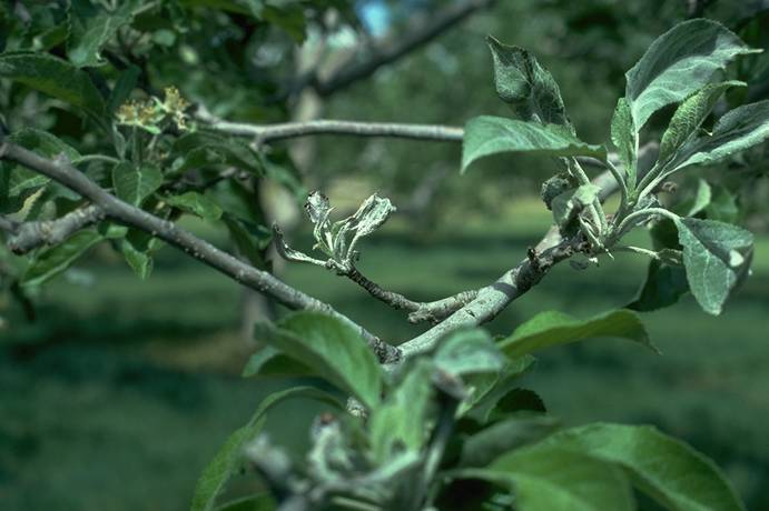
Powdery mildew-infected apple shoot. Note distorted leaves and white growth on leaves and along the shoot.
(Alan L. Jones, Michigan State University and Turner B. Sutton, North Carolina State University)
Powdery_Mildew_2
Powdery Mildew 2
White growth of powdery mildew fungus on apple leaves emerging from overwintered infected buds.
(Alan L. Jones, Michigan State University and Turner B. Sutton, North Carolina State University)

Powdery_Mildew_3
Powdery Mildew 3
Fruit showing net russeting from powdery mildew infection.
(Alan L. Jones, Michigan State University and Turner B. Sutton, North Carolina State University)
Powdery_mildew_apple_shoot_infection
Powdery mildew apple shoot infection (H. J. Larsen; Colorado State University)
Powdery_mildew_on_apple_2nd_growth_flush
Powdery mildew on apple 2nd growth flush (N. S. Luepschen; Colorado State University)
Powdery_mildew_pear_fruit_infection
Powdery mildew pear fruit infection (N. S. Luepschen; Colorado State University)
Powdery_mildew_pear_shoot_infection
Powdery mildew pear shoot infection (N. S. Luepschen; Colorado State University)
Powdery_mildew_russet_on_Jonathan_apple_fruit_(right_-_healthy)
Powdery mildew russet on Jonathan apple fruit (right - healthy) (N. S. Luepschen; Colorado State University)
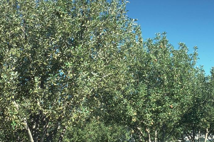
Powdery_mildew_vs_non_mildewed_apple_trees
Powdery mildew vs non mildewed apple trees (N. S. Luepschen; Colorado State University)
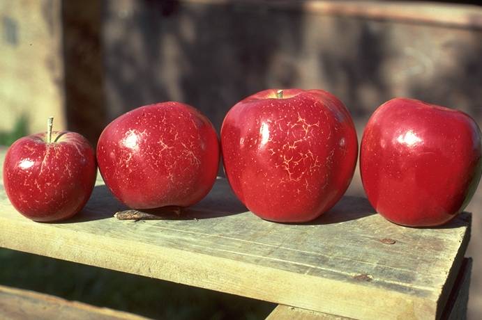
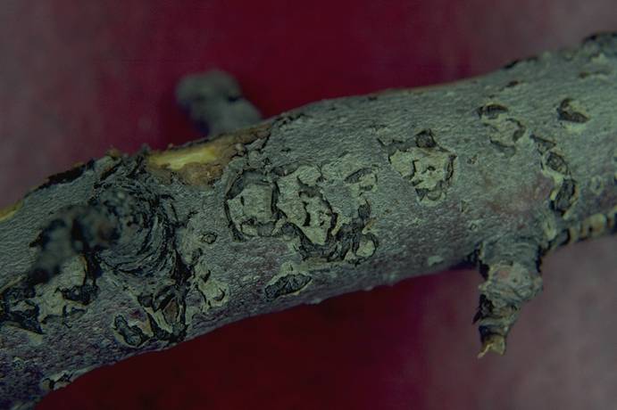
Red_delicious_apple,_blister_bark_(virus)_symptoms
Red delicious apple, blister bark (virus) symptoms (N. S. Luepschen; Colorado State University)
Red_delicious_apple,_dapple_apple_(viroid)_fruit_symptoms
Red delicious apple, dapple apple (viroid) fruit symptoms (H. J. Larsen; Colorado State University)
Red_delicious_apple,_flat_apple_(cherry_raspleaf_virus)_fruit_symptoms
Red delicious apple, flat apple (cherry raspleaf virus) fruit symptoms (H. J. Larsen; Colorado State University)
Rome_apple_blue_mold
Rome apple blue mold (N. S. Luepschen; Colorado State University)
Rome_apple_fire_blight_infection_on_scaffold
Rome apple fire blight infection on scaffold (N. S. Luepschen; Colorado State University)
Rome_apple_sunscald_+_bridge_graft
Rome apple sunscald + bridge graft (C. R. Ure; Colorado State University)
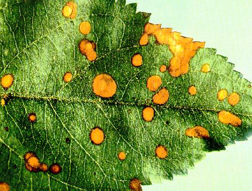
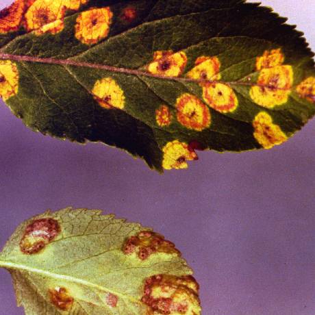
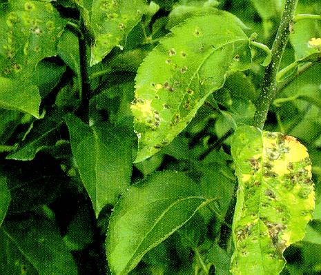
Rust_1
Rust 1
Necrotic spots with orange margins on apple leaves caused by cedar-apple rust.
(Alan L. Jones, Michigan State University and Turner B. Sutton, North Carolina State University)
Rust_2
Rust 2
Micrograph of a pycnium of cedar-apple rust fungus with a few periphyses extended and some pycniospores. Nectar drops were removed during preparation.
(Alan L. Jones, Michigan State University and Turner B. Sutton, North Carolina State University)
Rust_3
Rust 3
Cross-section of an apple leaf showing young aecia "cluster-cups" of the cedar-apple rust fungus on the underside of the leaf.
(Alan L. Jones, Michigan State University and Turner B. Sutton, North Carolina State University)
Rust_4
Rust 4
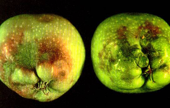
Distortion of calyx end and red discoloration of Delicious apple caused by quince rust.
(Alan L. Jones, Michigan State University and Turner B. Sutton, North Carolina State University)
Rust_5
Rust 5
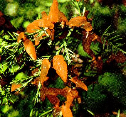
Tiny cedar-apple rust galls with gelatinous spore horns in axils of cedar leaves.
(Alan L. Jones, Michigan State University and Turner B. Sutton, North Carolina State University)
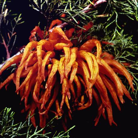
Rust_6
Rust 6
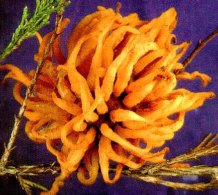
Cedar-apple rust gall on cedar with gelatinous spore horns fully extended.
(Alan L. Jones, Michigan State University and Turner B. Sutton, North Carolina State University)
Rust_7
Rust 7
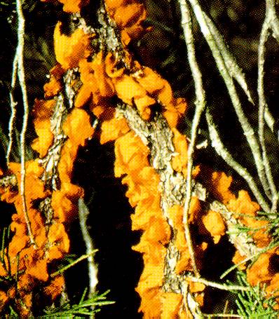
Quince rust canker on cedar with gelatinous spore horns fully extended.
(Alan L. Jones, Michigan State University and Turner B. Sutton, North Carolina State University)
Rust_8a
Rust 8a
Micrograph of a gelatinous spore horn from a cedar-apple rust fall. Teliospores of the cedar-apple rust fungus with slight constriction at the crosswall and single teliospore bearing promycelium (left) and formation of secondary basidiospore on a pointed sterigmata (right). Other basidiospores can be seen on the left.
(Alan L. Jones, Michigan State University and Turner B. Sutton, North Carolina State University)

Rust_8b
Rust 8b
Micrograph of a gelatinous spore horn from a cedar-apple rust fall. Teliospores of the cedar-apple rust fungus with slight constriction at the crosswall and single teliospore bearing promycelium (left) and formation of secondary basidiospore on a pointed sterigmata (right). Other basidiospores can be seen on the left.
(Alan L. Jones, Michigan State University and Turner B. Sutton, North Carolina State University)

Scab_on_Fruit_
Scab on Fruit
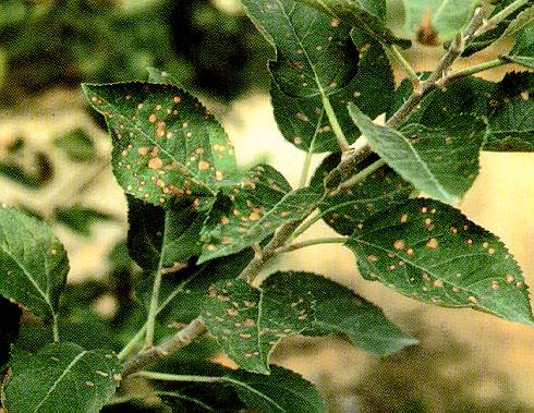
Scab_on_Young_Fruit_and_Leaf
Scab on Young Fruit and Leaf
Slime_flux_on_black_Jonathan_apple
Slime flux on black Jonathan apple (N. S. Luepschen; Colorado State University)
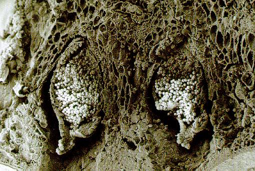
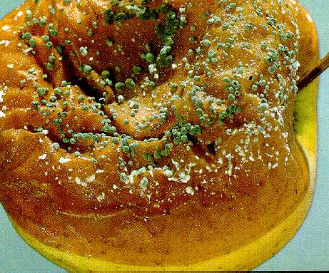
Soft_Rot/Blue_Mold_1
Soft Rot/Blue Mold 1
Golden Delicious apple with soft rot/blue mold. Note cushions of blue-green fungus growth on surface lesion.
(Alan L. Jones, Michigan State University and Turner B. Sutton, North Carolina State University)
Soft_Rot/Blue_Mold_2
Soft Rot/Blue Mold 2
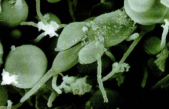
Conidiosphores of the soft rot pathogen Penicillium expansum. Note one-celled ovoid conidia produced on brush-like structures.
(Alan L. Jones, Michigan State University and Turner B. Sutton, North Carolina State University)
Sooty_Bloch_and_Flyspeck_1
Sooty Bloch and Flyspeck 1
Mycelial type of sooty blotch and a few flyspeck lesions on the surface of apple.
(Alan L. Jones, Michigan State University and Turner B. Sutton, North Carolina State University)
Sooty_Bloch_and_Flyspeck_2
Sooty Bloch and Flyspeck 2
Flyspeck lesions (groups of black, shiny dots) on the surface of Rome Beauty apple.
(Alan L. Jones, Michigan State University and Turner B. Sutton, North Carolina State University)
Sooty_Blotch_and_Flyspeck
Sooty Blotch and Flyspeck
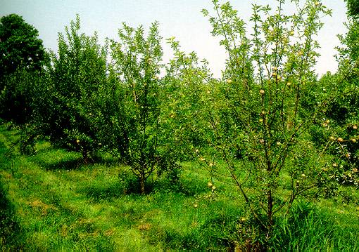
Southern_Blight_1
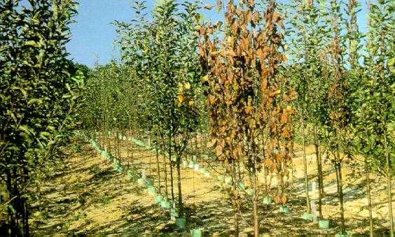
Southern Blight 1
Trees in a nursery row with dieback due to infection at soil line by the southern blight pathogens.
(Alan L. Jones, Michigan State University and Turner B. Sutton, North Carolina State University)
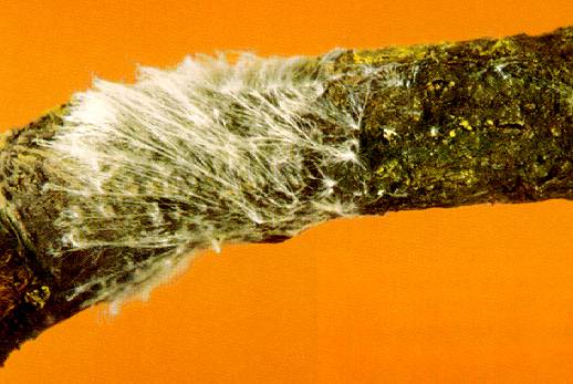
Southern_Blight_2
Southern Blight 2
White, web-like mycelium of Sclerotium rolfsii. Light brown to yellow, round sclerotia often form in the mycelial mat.
(Alan L. Jones, Michigan State University and Turner B. Sutton, North Carolina State University)
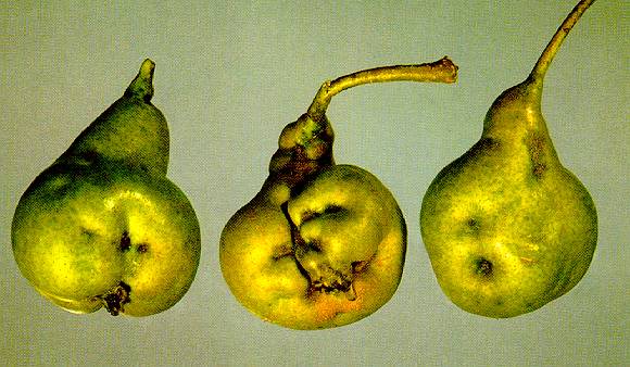
Stony_Pit_of_Pear
Stony Pit of Pear
Bosc pear fruit severely gnarled from stony pit.
(Alan L. Jones, Michigan State University and Turner B. Sutton, North Carolina State University)
Sulfur_burn_to_apple_foliage
Sulfur burn to apple foliage (N. S. Luepschen; Colorado State University)
Sulfur_burn_to_apple_foliage
Sulfur burn to apple foliage (N. S. Luepschen; Colorado State University)
Sulfur_burn_to_apple_fruit_and_foliage
Sulfur burn to apple fruit and foliage (N. S. Luepschen; Colorado State University)
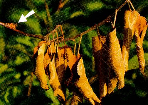
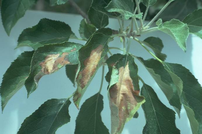
Thread_Blight
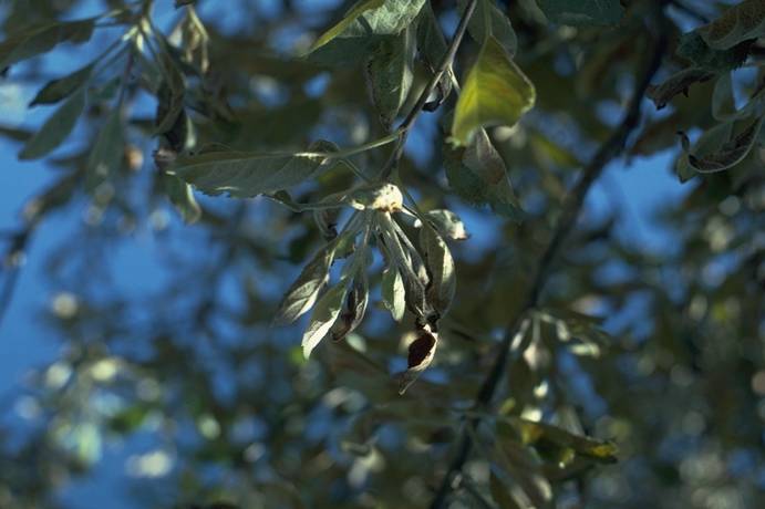
Thread Blight
Apple branch affected with thread blight showing dead leaves still attached.
(Alan L. Jones, Michigan State University and Turner B. Sutton, North Carolina State University)
Union_necrosis_(TmRS_virus)_of_apple
Union necrosis (TmRS virus) of apple (H. J. Larsen; Colorado State University)
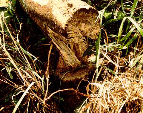
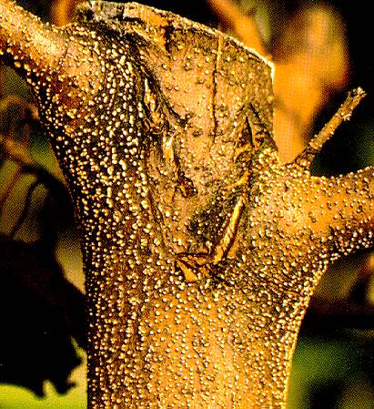
White_Root_Rot
White Root Rot
White mycelium of the white root rot fungus growing over the surface of the trunk.
(Alan L. Jones, Michigan State University and Turner B. Sutton, North Carolina State University)
White_Rot_1
White Rot 1
Early stages of white or bitter rot on fruit.
(Alan L. Jones, Michigan State University and Turner B. Sutton, North Carolina State University)
White_Rot_2
White Rot 2
Several examples of white rot on Golden Delicious fruit. Apple on lower left is infected with black rot.
(Alan L. Jones, Michigan State University and Turner B. Sutton, North Carolina State University)
White_Rot_3
White Rot 3
Internal fruit symptoms of bitter rot (left) and apple white rot (right). Decay from white rot (but not bitter rot) usually reached and surrounds the core.
(Alan L. Jones, Michigan State University and Turner B. Sutton, North Carolina State University)
White_Rot_4
White Rot 4
Reddish brown lesions on bark of Golden Delicious apple is the early symptom of white rot caused by Botryosphaeria dothidea.
(Alan L. Jones, Michigan State University and Turner B. Sutton, North Carolina State University)
White_Rot_5
White Rot 5
Exudation of liquid from a blister in an active white rot canker.
(Alan L. Jones, Michigan State University and Turner B. Sutton, North Carolina State University)
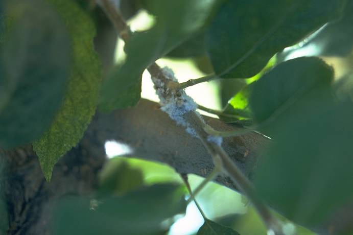
Woolly_aphids_on_apple
Woolly aphids on apple (N. S. Luepschen; Colorado State University)
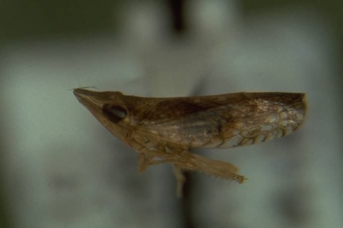
X_disease_vector_adult,_Fieberiella_florii
X disease vector adult, Fieberiella florii (C. L. Parish; Colorado State University)
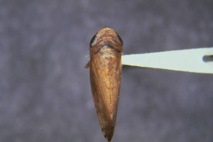
X_disease_vector_adult,_Fieberiella_florii
X disease vector adult, Fieberiella florii (C. L. Parish; Colorado State University)
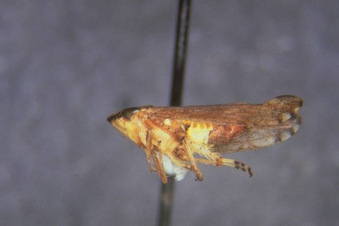
X_disease_vector_adult,_Scaphytopius_acutus
X disease vector adult, Scaphytopius acutus (C. L. Parish; Colorado State University)
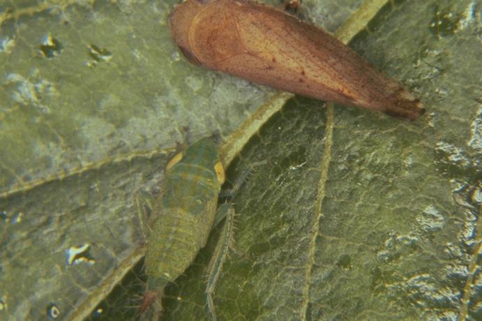
X_disease_vector_adult_&_nymph,_Fieberiella_florii
X disease vector adult & nymph, Fieberiella florii (C. L. Parish; Colorado State University)
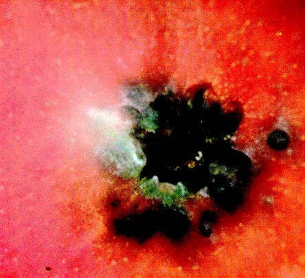
X-Spot
X-Spot
X-spot on the calyx end of apple fruit.
(Alan L. Jones, Michigan State University and Turner B. Sutton, North Carolina State University)
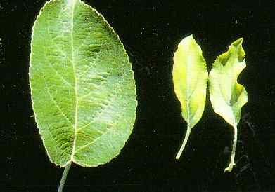
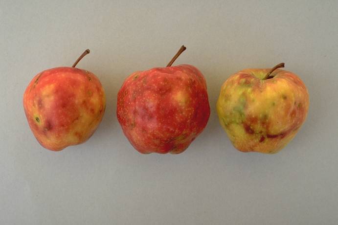
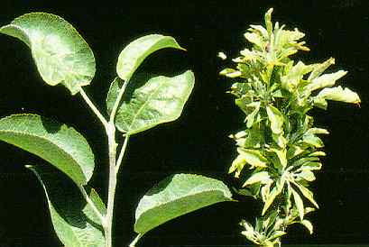
Zinc_deficiency_1
Zinc deficiency (right), healthy leaves (left)
(S. V. Thomson, Utah State University)
Zinc_deficiency_2
Zinc deficiency (right), healthy leaf (left) (S. V. Thomson, Utah State University)